Volume 24, Number 3—March 2018
Dispatch
Invasive Infections Caused by Nannizziopsis spp. Molds in Immunocompromised Patients
Figure

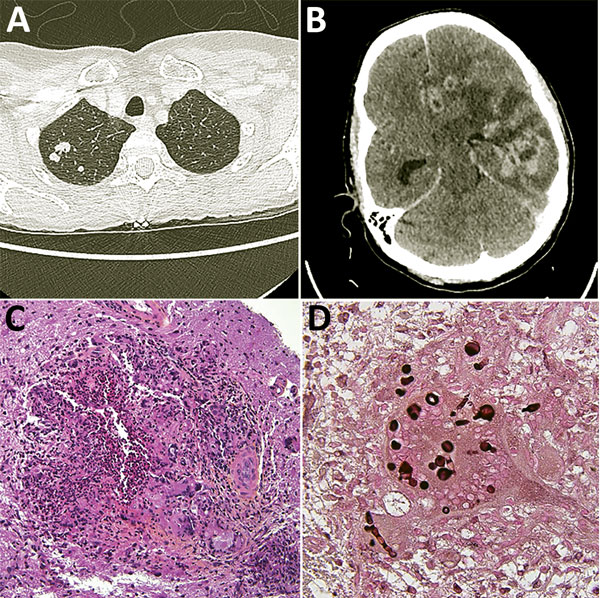
Figure. Diagnostic testing of a 52-year-old woman from France living in Mali who had Nannizziopsis spp. fungal infection. A) Thoracic-abdominal-pelvic scan shows pseudo-nodular lesions in the apex of the right lung, of which one is excavated. B) Cerebral computed tomography scan shows contrast enhancement on several hemispheric nodules on the left and in frontal, parietal, and temporal regions, responsible for large surrounding edema and compression of the left lateral ventricle. The median line is deviated to the right with a subfalcorial herniation. C) Hematoxylin-eosin-saffron stain of brain biopsy containing mononuclear inflammatory infiltrates; giant cell granulomas; histiocytes, sometimes with an epithelioid appearance; and neutrophils (original magnification ×200). D) Grocott stain showing thick bulbous mycelial filaments in the cytoplasm of certain giant cells/histiocytes (original magnification ×600). Round shapes correspond to cross-sections of bulbous territories.