Volume 24, Number 5—May 2018
Dispatch
Heartland Virus and Hemophagocytic Lymphohistiocytosis in Immunocompromised Patient, Missouri, USA
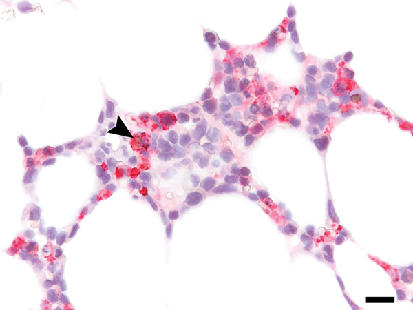
Figure 2

Figure 2. Immunohistochemistry of bone marrow from immunocompromised patient infected with Heartland virus (HRTV), Missouri, USA. Testing of biopsied sample from post–symptom onset day 11 shows extensive positive staining for HRTV antigen, including erythrophagocytosis by an HRTV-antigen–positive cell (arrowhead). Scale bar indicates 20μm.
Page created: April 17, 2018
Page updated: April 17, 2018
Page reviewed: April 17, 2018
The conclusions, findings, and opinions expressed by authors contributing to this journal do not necessarily reflect the official position of the U.S. Department of Health and Human Services, the Public Health Service, the Centers for Disease Control and Prevention, or the authors' affiliated institutions. Use of trade names is for identification only and does not imply endorsement by any of the groups named above.