Volume 24, Number 6—June 2018
Research
Novel Parvovirus Related to Primate Bufaviruses in Dogs
Figure 1
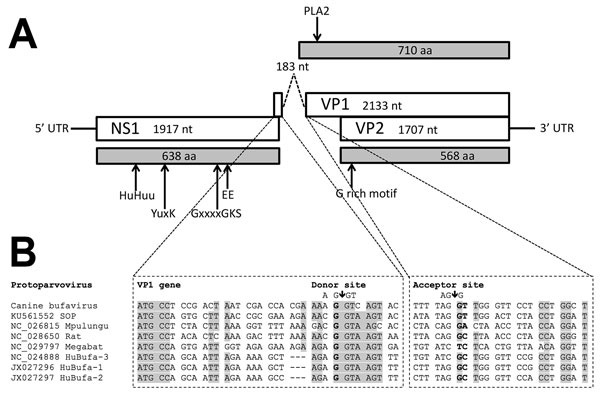
Figure 1. Genome organization of canine bufavirus. A) Positions of the conserved helicase Walker A (GxxxxGKS), Walker B (EE), and replication initiator motifs (HuHuu and YuxK) in NS1 and of the phospholipase A2 (PLA2) and glycine-rich region (G-rich) in VP1 and VP2. B) Putative splicing mechanism in the VP1 gene of canine bufavirus, human bufaviruses, and other protoparvoviruses. Two potential splice sites are a potential donor site (AG↓GT) at nt 1931 and an acceptor site (AG↓G) at nt 2115. The putative VP1 sequence starts with ATG at the end of ORF1 at nt 1906 upstream of the splice donor site at nt 1931. Gray shading indicates strictly and highly conserved bases. GenBank accession numbers are provided for reference sequences. NS, nonstructural; UTR, untranslated region; VP, viral capsid protein.