Volume 24, Number 7—July 2018
Research
Diversity of Influenza A(H5N1) Viruses in Infected Humans, Northern Vietnam, 2004–2010
Figure 2

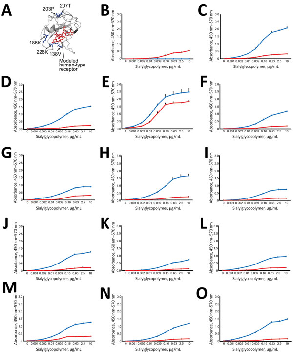
Figure 2. Effect of amino acid variations in hemagglutinin (HA) on influenza virus receptor–binding specificities in influenza A(H5N1) virus isolates from humans, northern Vietnam, 2004–2010. A) Localization of selected amino acid variations detected in clinical samples. The detected HA changes were mapped onto the receptor-binding domain (aa positions 117–265) of a monomer of A/VN1203/2004 (H5N1) HA (Protein Data Bank accession no. 2FK0). Red indicates modeled human-type receptor; blue indicates positions of amino acid variations on the receptor-binding domain. B–O) Receptor-binding specificities of HA variants. Direct binding of virus to α2,3-linked (blue) or α2,6-linked (red) sialylglycopolymers was assessed. Shown is the mean receptor-binding specificity + SDs of triplicates of a single experiment. If 2 isolate numbers are listed (e.g., CA04/UT3040I/II), we tested the major sequence variant (which is identical between the 2 samples: B) CA04/K173; C) CA04/UT3040I/II-HA; D) CA04/UT3040I-HA-138V; E) CA04/UT3040II-HA-186K; F) CA04/UT31312III-HA; G) CA04/UT31312III-HA-186K; H) CA04/UT31312III-HA-203P-to-L (the proline residue introduced at position 203 of UT31312III HA mutated to leucine); I) CA04/UT31394II-HA; J) CA04/UT31394II-HA-207T; K) CA04/UT36250I/II-HA; L) CA04/UT36250I/II-HA-138V; M) CA04/UT36250II-HA-226K; N) CA04/UT36282I/II-HA; O) CA04/UT36282I/II-HA-138V/A (after introduction of valine at position 138 of UT36282I/II HA, we detected both valine and alanine at this position).
1These authors contributed equally to this article.