Volume 24, Number 8—August 2018
Dispatch
Anncaliia algerae Microsporidial Myositis, New South Wales, Australia
Gaurav Sutrave, Adam Maundrell, Caitlin Keighley, Zoe Jennings, Susan Brammah, Min-Xia Wang, Roger Pamphlett, Cameron E. Webb, Damien Stark, Helen Englert, David Gottlieb, Ian Bilmon, and Matthew R. Watts
Figure 1
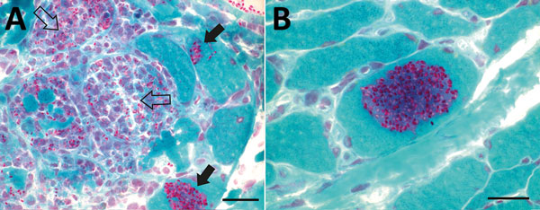
Figure 1. Light micrographs of Gomori trichrome–stained frozen sections of vastus lateralis muscle from a 66-year-old man with Anncaliia algerae microsporidial myositis, New South Wales, Australia. A) Necrotising myositis with red-stained, ovoid spores in green-staining viable myocytes (solid arrows) and within macrophages invading necrotic myocytes (open arrows). B) A cluster of red stained, 2–3 µm spores within a viable myocyte. Scale bars indicate 25 µm.
Page created: July 18, 2018
Page updated: July 18, 2018
Page reviewed: July 18, 2018
The conclusions, findings, and opinions expressed by authors contributing to this journal do not necessarily reflect the official position of the U.S. Department of Health and Human Services, the Public Health Service, the Centers for Disease Control and Prevention, or the authors' affiliated institutions. Use of trade names is for identification only and does not imply endorsement by any of the groups named above.