Volume 25, Number 1—January 2019
Research Letter
Severe Disseminated Infection with Emerging Lineage of Methicillin-Sensitive Staphylococcus aureus
Paul Jewell , Luke Dixon, Aran Singanayagam, Rohma Ghani, Ernie Wong, Meg Coleman, Bruno Pichon, Angela Kearns, Georgina Russell, and James Hatcher
Figure
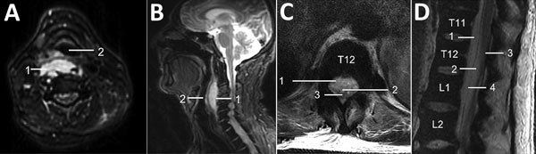
Figure. Magnetic resonance imaging of a 60-year-old immunocompetent man with methicillin-resistant Staphylococcus aureus clonal complex 398 infection. A, B) Axial (A) and sagittal (B) T2-weighted fat-suppressed sequences of the cervical spine demonstrate a retropharyngeal abscess (1) that moderately anteriorly displaces and mildly effaces the hypopharynx (2). C, D) Axial (C) and sagittal (D) T2-weighted MRI sequences of the thoracolumbar spine (T11–L2 vertebra levels labeled) demonstrate a large ventral, combined epidural (1) and subdural (2) spinal collection that displaces the conus medullaris (3) dorsally. Note the dura mater (4) on the sagittal sequence, which delineates the theca and separates the epidural and subdural spaces.
Page created: December 18, 2018
Page updated: December 18, 2018
Page reviewed: December 18, 2018
The conclusions, findings, and opinions expressed by authors contributing to this journal do not necessarily reflect the official position of the U.S. Department of Health and Human Services, the Public Health Service, the Centers for Disease Control and Prevention, or the authors' affiliated institutions. Use of trade names is for identification only and does not imply endorsement by any of the groups named above.