Volume 25, Number 10—October 2019
Dispatch
Possible Prognostic Value of Serial Brain MRIs in Powassan Virus Encephalitis
Joshua Allgaier , Ryan Quarles, and Daniel Skiest
Figure
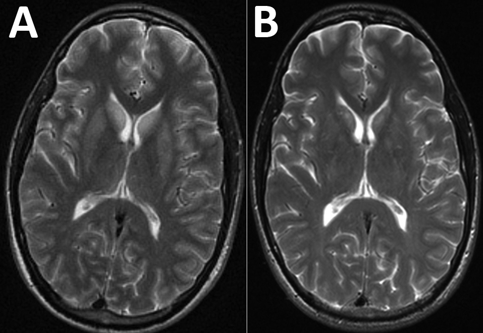
Figure. Magnetic resonance imaging (MRI) of the brain of a patient with encephalitis caused by Powassan virus, Massachusetts, USA, 2017. A) Initial brain MRI showing high T2 signal abnormality in the bilateral caudate and putamen. B) Noticeable improvement on repeat brain MRI 2 weeks later.
Page created: September 17, 2019
Page updated: September 17, 2019
Page reviewed: September 17, 2019
The conclusions, findings, and opinions expressed by authors contributing to this journal do not necessarily reflect the official position of the U.S. Department of Health and Human Services, the Public Health Service, the Centers for Disease Control and Prevention, or the authors' affiliated institutions. Use of trade names is for identification only and does not imply endorsement by any of the groups named above.