Volume 25, Number 11—November 2019
Research
Nasopharyngeal Pneumococcal Density during Asymptomatic Respiratory Virus Infection and Risk for Subsequent Acute Respiratory Illness
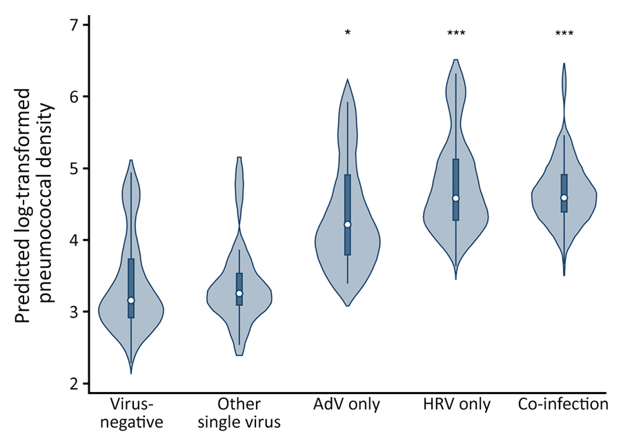
Figure 2

Figure 2. Violin plots of predicted log10-transformed pneumococcal colonization densities by detection of specific viruses among children <3 years of age, Respiratory Infections in Andean Peruvian Children study, May 2009–September 2011. Predicted densities were estimated from the final multivariable linear quantile mixed effects model. Circles indicate median densities, boxes represent interquartile range, lines extend through the upper and lower adjacent values, and the density plot width indicates the predicted frequency of observations. *p<0.05; ***p<0.001. AdV, adenovirus; HRV, rhinovirus.
Page created: October 15, 2019
Page updated: October 15, 2019
Page reviewed: October 15, 2019
The conclusions, findings, and opinions expressed by authors contributing to this journal do not necessarily reflect the official position of the U.S. Department of Health and Human Services, the Public Health Service, the Centers for Disease Control and Prevention, or the authors' affiliated institutions. Use of trade names is for identification only and does not imply endorsement by any of the groups named above.