Synopses
Vaccine-Derived Poliovirus Infection among Patients with Primary Immunodeficiency and Effect of Patient Screening on Disease Outcomes, Iran [PDF - 894 KB - 8 pages]
Patients with immunodeficiency-associated vaccine-derived poliovirus (iVDPV) are potential poliovirus reservoirs in the posteradication era that might reintroduce polioviruses into the community. We update the iVDPV registry in Iran by reporting 9 new patients. In addition to national acute flaccid paralysis surveillance, cases were identified by screening nonparalyzed primary immunodeficiency (PID) patients. Overall, 23 iVDPV patients have been identified since 1995. Seven patients (30%) never had paralysis. Poliovirus screening accelerated the iVDPV detection rate in Iran after 2014.The iVDPV infection rate among nonparalyzed patients with adaptive PID was 3.1% (7/224), several folds higher than previous estimates. Severe combined immunodeficiency patients had the highest risk for asymptomatic infection (28.6%) compared with other PIDs. iVDPV2 emergence has decreased after the switch from trivalent to bivalent oral poliovirus vaccine in 2016. However, emergence of iVDPV1 and iVDPV3 continued. Poliovirus screening in PID patients is an essential step in the endgame of polio eradication.
| EID | Shaghaghi M, Shahmahmoodi S, Nili A, Abolhassani H, Madani S, Nejati A, et al. Vaccine-Derived Poliovirus Infection among Patients with Primary Immunodeficiency and Effect of Patient Screening on Disease Outcomes, Iran. Emerg Infect Dis. 2019;25(11):2005-2012. https://doi.org/10.3201/eid2511.190540 |
|---|---|
| AMA | Shaghaghi M, Shahmahmoodi S, Nili A, et al. Vaccine-Derived Poliovirus Infection among Patients with Primary Immunodeficiency and Effect of Patient Screening on Disease Outcomes, Iran. Emerging Infectious Diseases. 2019;25(11):2005-2012. doi:10.3201/eid2511.190540. |
| APA | Shaghaghi, M., Shahmahmoodi, S., Nili, A., Abolhassani, H., Madani, S., Nejati, A....Aghamohammadi, A. (2019). Vaccine-Derived Poliovirus Infection among Patients with Primary Immunodeficiency and Effect of Patient Screening on Disease Outcomes, Iran. Emerging Infectious Diseases, 25(11), 2005-2012. https://doi.org/10.3201/eid2511.190540. |
Research
Comparison of Whole-Genome Sequences of Legionella pneumophila in Tap Water and in Clinical Strains, Flint, Michigan, USA, 2016 [PDF - 1.23 MB - 8 pages]
During the water crisis in Flint, Michigan, USA (2014–2015), 2 outbreaks of Legionnaires’ disease occurred in Genesee County, Michigan. We compared whole-genome sequences of 10 clinical Legionella pneumophila isolates submitted to a laboratory in Genesee County during the second outbreak with 103 water isolates collected the following year. We documented a genetically diverse range of L. pneumophila strains across clinical and water isolates. Isolates belonging to 1 clade (3 clinical isolates, 3 water isolates from a Flint hospital, 1 water isolate from a Flint residence, and the reference Paris strain) had a high degree of similarity (2–1,062 single-nucleotide polymorphisms), all L. pneumophila sequence type 1, serogroup 1. Serogroup 6 isolates belonging to sequence type 2518 were widespread in Flint hospital water samples but bore no resemblance to available clinical isolates. L. pneumophila strains in Flint tap water after the outbreaks were diverse and similar to some disease-causing strains.
| EID | Garner E, Brown CL, Schwake D, Rhoads WJ, Arango-Argoty G, Zhang L, et al. Comparison of Whole-Genome Sequences of Legionella pneumophila in Tap Water and in Clinical Strains, Flint, Michigan, USA, 2016. Emerg Infect Dis. 2019;25(11):2013-2020. https://doi.org/10.3201/eid2511.181032 |
|---|---|
| AMA | Garner E, Brown CL, Schwake D, et al. Comparison of Whole-Genome Sequences of Legionella pneumophila in Tap Water and in Clinical Strains, Flint, Michigan, USA, 2016. Emerging Infectious Diseases. 2019;25(11):2013-2020. doi:10.3201/eid2511.181032. |
| APA | Garner, E., Brown, C. L., Schwake, D., Rhoads, W. J., Arango-Argoty, G., Zhang, L....Pruden, A. (2019). Comparison of Whole-Genome Sequences of Legionella pneumophila in Tap Water and in Clinical Strains, Flint, Michigan, USA, 2016. Emerging Infectious Diseases, 25(11), 2013-2020. https://doi.org/10.3201/eid2511.181032. |
Clinical and Molecular Epidemiology of Invasive Group B Streptococcus Disease among Infants, China [PDF - 1.87 MB - 10 pages]
Invasive group B Streptococcus (GBS) remains a leading cause of illness and death among infants globally. We conducted prospective and retrospective laboratory-based surveillance of GBS-positive cultures from infants <3 months of age in 18 hospitals across China during January 1, 2015–December 31, 2017. The overall incidence of GBS was 0.31 (95% CI 0.27–0.36) cases/1,000 live births; incidence was 0–0.76 cases/1,000 live births across participating hospitals. The case-fatality rate was 2.3%. We estimated 13,604 cases of GBS and 1,142 GBS–associated deaths in infants <90 days of age annually in China. GBS isolates were most commonly serotype III (61.5%) and clonal complex 17 (40.6%). Enhanced active surveillance and implementation of preventive strategies, such as maternal GBS vaccination, warrants further investigation in China to help prevent these infections.
| EID | Ji W, Liu H, Madhi SA, Cunnington M, Zhang Z, Dangor Z, et al. Clinical and Molecular Epidemiology of Invasive Group B Streptococcus Disease among Infants, China. Emerg Infect Dis. 2019;25(11):2021-2030. https://doi.org/10.3201/eid2511.181647 |
|---|---|
| AMA | Ji W, Liu H, Madhi SA, et al. Clinical and Molecular Epidemiology of Invasive Group B Streptococcus Disease among Infants, China. Emerging Infectious Diseases. 2019;25(11):2021-2030. doi:10.3201/eid2511.181647. |
| APA | Ji, W., Liu, H., Madhi, S. A., Cunnington, M., Zhang, Z., Dangor, Z....Fang, Y. (2019). Clinical and Molecular Epidemiology of Invasive Group B Streptococcus Disease among Infants, China. Emerging Infectious Diseases, 25(11), 2021-2030. https://doi.org/10.3201/eid2511.181647. |
Seasonal Influenza and Avian Influenza A(H5N1) Virus Surveillance among Inpatients and Outpatients, East Jakarta, Indonesia, 2011–2014 [PDF - 1.04 MB - 9 pages]
During October 2011–September 2014, we screened respiratory specimens for seasonal and avian influenza A(H5N1) virus infections among outpatients with influenza-like illness and inpatients with severe acute respiratory infection (SARI) in East Jakarta, an Indonesia district with high incidence of H5N1 virus infection among poultry. In total, 31% (1,875/6,008) of influenza-like illness case-patients and 15% (571/3,811) of SARI case-patients tested positive for influenza virus. Influenza A(H1N1)pdm09, influenza A(H3N2), and influenza B virus infections were detected in all 3 years, and the epidemic season extended from November through May. Although 28% (2,810/10,135) of case-patients reported exposure to poultry, only 1 SARI case-patient with an H5N1 virus infection was detected. Therefore, targeted screening among case-patients with high-risk poultry exposures (e.g., a recent visit to a live bird market or close proximity to sick or dead poultry) may be a more efficient routine surveillance strategy for H5N1 virus in these types of settings.
| EID | Lafond KE, Praptiningsih CY, Mangiri A, Syarif M, Triada R, Mulyadi E, et al. Seasonal Influenza and Avian Influenza A(H5N1) Virus Surveillance among Inpatients and Outpatients, East Jakarta, Indonesia, 2011–2014. Emerg Infect Dis. 2019;25(11):2031-2039. https://doi.org/10.3201/eid2511.181844 |
|---|---|
| AMA | Lafond KE, Praptiningsih CY, Mangiri A, et al. Seasonal Influenza and Avian Influenza A(H5N1) Virus Surveillance among Inpatients and Outpatients, East Jakarta, Indonesia, 2011–2014. Emerging Infectious Diseases. 2019;25(11):2031-2039. doi:10.3201/eid2511.181844. |
| APA | Lafond, K. E., Praptiningsih, C. Y., Mangiri, A., Syarif, M., Triada, R., Mulyadi, E....Iuliano, A. (2019). Seasonal Influenza and Avian Influenza A(H5N1) Virus Surveillance among Inpatients and Outpatients, East Jakarta, Indonesia, 2011–2014. Emerging Infectious Diseases, 25(11), 2031-2039. https://doi.org/10.3201/eid2511.181844. |
Nasopharyngeal Pneumococcal Density during Asymptomatic Respiratory Virus Infection and Risk for Subsequent Acute Respiratory Illness [PDF - 928 KB - 8 pages]
Increased nasopharyngeal pneumococcal (Streptococcus pneumoniae) colonization density has been associated with invasive pneumococcal disease, but factors that increase pneumococcal density are poorly understood. We evaluated pneumococcal densities in nasopharyngeal samples from asymptomatic young children from Peru and their association with subsequent acute respiratory illness (ARI). Total pneumococcal densities (encompassing all present serotypes) during asymptomatic periods were significantly higher when a respiratory virus was detected versus when no virus was detected (p<0.001). In adjusted analyses, increased pneumococcal density was significantly associated with the risk for a subsequent ARI (p<0.001), whereas asymptomatic viral detection alone was associated with lower risk for subsequent ARI. These findings suggest that interactions between viruses and pneumococci in the nasopharynx during asymptomatic periods might have a role in onset of subsequent ARI. The mechanisms for these interactions, along with other potentially associated host and environmental factors, and their role in ARI pathogenesis and pneumococcal transmission require further elucidation.
| EID | Howard LM, Zhu Y, Griffin MR, Edwards KM, Williams JV, Gil AI, et al. Nasopharyngeal Pneumococcal Density during Asymptomatic Respiratory Virus Infection and Risk for Subsequent Acute Respiratory Illness. Emerg Infect Dis. 2019;25(11):2040-2047. https://doi.org/10.3201/eid2511.190157 |
|---|---|
| AMA | Howard LM, Zhu Y, Griffin MR, et al. Nasopharyngeal Pneumococcal Density during Asymptomatic Respiratory Virus Infection and Risk for Subsequent Acute Respiratory Illness. Emerging Infectious Diseases. 2019;25(11):2040-2047. doi:10.3201/eid2511.190157. |
| APA | Howard, L. M., Zhu, Y., Griffin, M. R., Edwards, K. M., Williams, J. V., Gil, A. I....Grijalva, C. G. (2019). Nasopharyngeal Pneumococcal Density during Asymptomatic Respiratory Virus Infection and Risk for Subsequent Acute Respiratory Illness. Emerging Infectious Diseases, 25(11), 2040-2047. https://doi.org/10.3201/eid2511.190157. |
Rare Detection of Bordetella pertussis Pertactin-Deficient Strains in Argentina [PDF - 1.18 MB - 7 pages]
Pertussis resurgence had been attributed to waning vaccine immunity and Bordetella pertussis adaptation to escape vaccine-induced immunity. Circulating bacteria differ genotypically from strains used in production of pertussis vaccine. Pertactin-deficient strains are highly prevalent in countries that use acellular vaccine (aP), suggesting strong aP-imposed selection of circulating bacteria. To corroborate this hypothesis, systematic studies on pertactin prevalence of infection in countries using whole-cell vaccine are needed. We provide pertussis epidemiologic data and molecular characterization of B. pertussis isolates from Buenos Aires, Argentina, during 2000–2017. This area used primary vaccination with whole-cell vaccine. Since 2002, pertussis case incidences increased at regular 4-year outbreaks; most cases were in infants <1 year of age. Of the B. pertussis isolates analyzed, 90.6% (317/350) contained the ptxP3-ptxA1-prn2-fim3-2 allelic profile. Immunoblotting and sequencing techniques detected only the 2 pertactin-deficient isolates. The low prevalence of pertactin-deficient strains in Argentina suggests that loss of pertactin gene expression might be driven by aP vaccine.
| EID | Carriquiriborde F, Regidor V, Aispuro PM, Magali G, Bartel E, Bottero D, et al. Rare Detection of Bordetella pertussis Pertactin-Deficient Strains in Argentina. Emerg Infect Dis. 2019;25(11):2048-2054. https://doi.org/10.3201/eid2511.190329 |
|---|---|
| AMA | Carriquiriborde F, Regidor V, Aispuro PM, et al. Rare Detection of Bordetella pertussis Pertactin-Deficient Strains in Argentina. Emerging Infectious Diseases. 2019;25(11):2048-2054. doi:10.3201/eid2511.190329. |
| APA | Carriquiriborde, F., Regidor, V., Aispuro, P. M., Magali, G., Bartel, E., Bottero, D....Hozbor, D. (2019). Rare Detection of Bordetella pertussis Pertactin-Deficient Strains in Argentina. Emerging Infectious Diseases, 25(11), 2048-2054. https://doi.org/10.3201/eid2511.190329. |
Molecular and Clinical Comparison of Enterovirus D68 Outbreaks among Hospitalized Children, Ohio, USA, 2014 and 2018 [PDF - 2.19 MB - 9 pages]
Enterovirus D68 (EV-D68) causes respiratory tract infections and neurologic manifestations. We compared the clinical manifestations from 2 EV-D68 outbreaks in 2014 and 2018 and a low-activity period in 2016 among hospitalized children in central Ohio, USA, and used PCR and sequencing to enable phylogenetic comparisons. During both outbreak periods, infected children had respiratory manifestations that led to an increase in hospital admissions for asthma. The 2018 EV-D68 outbreak appeared to be milder in terms of respiratory illness, as shown by lower rates of pediatric intensive care unit admission. However, the frequency of severe neurologic manifestations was higher in 2018 than in 2014. During the same period in 2016, we noted neither an increase in EV-D68 nor a significant increase in asthma-related admissions. Phylogenetic analyses showed that EV-D68 isolates from 2018 clustered differently within clade B than did isolates from 2014 and are perhaps associated with a different EV-D68 subclade.
| EID | Wang H, Diaz A, Moyer K, Mele-Casas M, Ara-Montojo M, Torrus I, et al. Molecular and Clinical Comparison of Enterovirus D68 Outbreaks among Hospitalized Children, Ohio, USA, 2014 and 2018. Emerg Infect Dis. 2019;25(11):2055-2063. https://doi.org/10.3201/eid2511.190973 |
|---|---|
| AMA | Wang H, Diaz A, Moyer K, et al. Molecular and Clinical Comparison of Enterovirus D68 Outbreaks among Hospitalized Children, Ohio, USA, 2014 and 2018. Emerging Infectious Diseases. 2019;25(11):2055-2063. doi:10.3201/eid2511.190973. |
| APA | Wang, H., Diaz, A., Moyer, K., Mele-Casas, M., Ara-Montojo, M., Torrus, I....Leber, A. L. (2019). Molecular and Clinical Comparison of Enterovirus D68 Outbreaks among Hospitalized Children, Ohio, USA, 2014 and 2018. Emerging Infectious Diseases, 25(11), 2055-2063. https://doi.org/10.3201/eid2511.190973. |
West Nile Virus (WNV) can result in clinically severe neurologic disease. There is no treatment for WNV infection, but administration of anti-WNV polyclonal human antibody has demonstrated efficacy in animal models. We compared Omr-IgG-am, an immunoglobulin product with high titers of anti-WNV antibody, with intravenous immunoglobulin (IVIG) and normal saline to assess safety and efficacy in patients with WNV neuroinvasive disease as part of a phase I/II, randomized, double-blind, multicenter study in North America. During 2003–2006, a total of 62 hospitalized patients were randomized to receive Omr-IgG-am, standard IVIG, or normal saline (3:1:1). The primary endpoint was medication safety. Secondary endpoints were morbidity and mortality, measured using 4 standardized assessments of cognitive and functional status. The death rate in the study population was 12.9%. No significant differences were found between groups receiving Omr-IgG-am compared with IVIG or saline for either the safety or efficacy endpoints.
| EID | Gnann JW, Agrawal A, Hart J, Buitrago M, Carson P, Hanfelt-Goade D, et al. Lack of Efficacy of High-Titered Immunoglobulin in Patients with West Nile Virus Central Nervous System Disease. Emerg Infect Dis. 2019;25(11):2064-2073. https://doi.org/10.3201/eid2511.190537 |
|---|---|
| AMA | Gnann JW, Agrawal A, Hart J, et al. Lack of Efficacy of High-Titered Immunoglobulin in Patients with West Nile Virus Central Nervous System Disease. Emerging Infectious Diseases. 2019;25(11):2064-2073. doi:10.3201/eid2511.190537. |
| APA | Gnann, J. W., Agrawal, A., Hart, J., Buitrago, M., Carson, P., Hanfelt-Goade, D....Whitley, R. J. (2019). Lack of Efficacy of High-Titered Immunoglobulin in Patients with West Nile Virus Central Nervous System Disease. Emerging Infectious Diseases, 25(11), 2064-2073. https://doi.org/10.3201/eid2511.190537. |
Serosurvey for Influenza D Virus Exposure in Cattle, United States, 2014–2015 [PDF - 1.50 MB - 7 pages]
Influenza D virus has been detected predominantly in cattle from several countries. In the United States, regional and state seropositive rates for influenza D have previously been reported, but little information exists to evaluate national seroprevalence. We performed a serosurveillance study with 1,992 bovine serum samples collected across the country in 2014 and 2015. We found a high overall seropositive rate of 77.5% nationally; regional rates varied from 47.7% to 84.6%. Samples from the Upper Midwest and Mountain West regions showed the highest seropositive rates. In addition, seropositive samples were found in 41 of the 42 states from which cattle originated, demonstrating that influenza D virus circulated widely in cattle during this period. The distribution of influenza D virus in cattle from the United States highlights the need for greater understanding about pathogenesis, epidemiology, and the implications for animal health.
| EID | Silveira S, Falkenberg SM, Kaplan BS, Crossley B, Ridpath JF, Bauermann FB, et al. Serosurvey for Influenza D Virus Exposure in Cattle, United States, 2014–2015. Emerg Infect Dis. 2019;25(11):2074-2080. https://doi.org/10.3201/eid2511.190253 |
|---|---|
| AMA | Silveira S, Falkenberg SM, Kaplan BS, et al. Serosurvey for Influenza D Virus Exposure in Cattle, United States, 2014–2015. Emerging Infectious Diseases. 2019;25(11):2074-2080. doi:10.3201/eid2511.190253. |
| APA | Silveira, S., Falkenberg, S. M., Kaplan, B. S., Crossley, B., Ridpath, J. F., Bauermann, F. B....Neill, J. D. (2019). Serosurvey for Influenza D Virus Exposure in Cattle, United States, 2014–2015. Emerging Infectious Diseases, 25(11), 2074-2080. https://doi.org/10.3201/eid2511.190253. |
Dispatches
High Prevalence of Mansonella ozzardi Infection in the Amazon Region, Ecuador [PDF - 848 KB - 3 pages]
We reviewed Giemsa-stained thick blood smears, obtained through the national malaria surveillance program in the Amazon region of Ecuador, by light microscopy for Mansonella spp. microfilariae. Of 2,756 slides examined, 566 (20.5%) were positive. Nested PCR confirmed that the microfilariae were those of M. ozzardi nematodes, indicating that this parasite is endemic to this region.
| EID | Calvopina M, Chiluisa-Guacho C, Toapanta A, Fonseca D, Villacres I. High Prevalence of Mansonella ozzardi Infection in the Amazon Region, Ecuador. Emerg Infect Dis. 2019;25(11):2081-2083. https://doi.org/10.3201/eid2511.181964 |
|---|---|
| AMA | Calvopina M, Chiluisa-Guacho C, Toapanta A, et al. High Prevalence of Mansonella ozzardi Infection in the Amazon Region, Ecuador. Emerging Infectious Diseases. 2019;25(11):2081-2083. doi:10.3201/eid2511.181964. |
| APA | Calvopina, M., Chiluisa-Guacho, C., Toapanta, A., Fonseca, D., & Villacres, I. (2019). High Prevalence of Mansonella ozzardi Infection in the Amazon Region, Ecuador. Emerging Infectious Diseases, 25(11), 2081-2083. https://doi.org/10.3201/eid2511.181964. |
Clinical REsearch During Outbreaks (CREDO) Training for Low- and Middle-Income Countries [PDF - 576 KB - 4 pages]
We describe a pilot of the Clinical REsearch During Outbreaks (CREDO) initiative, a training curriculum for researchers in epidemic-prone low- and middle-income countries who may respond to disease outbreaks. Participants reported improved confidence in their ability to conduct such research and overall satisfaction with the course structure, content, and training.
| EID | Kayem N, Rojek A, Denis E, Salam A, Reis A, Olliaro P, et al. Clinical REsearch During Outbreaks (CREDO) Training for Low- and Middle-Income Countries. Emerg Infect Dis. 2019;25(11):2084-2087. https://doi.org/10.3201/eid2511.180628 |
|---|---|
| AMA | Kayem N, Rojek A, Denis E, et al. Clinical REsearch During Outbreaks (CREDO) Training for Low- and Middle-Income Countries. Emerging Infectious Diseases. 2019;25(11):2084-2087. doi:10.3201/eid2511.180628. |
| APA | Kayem, N., Rojek, A., Denis, E., Salam, A., Reis, A., Olliaro, P....Horby, P. (2019). Clinical REsearch During Outbreaks (CREDO) Training for Low- and Middle-Income Countries. Emerging Infectious Diseases, 25(11), 2084-2087. https://doi.org/10.3201/eid2511.180628. |
Non-Leishmania Parasite in Fatal Visceral Leishmaniasis–Like Disease, Brazil [PDF - 5.89 MB - 5 pages]
Through whole-genome sequencing analysis, we identified non-Leishmania parasites isolated from a man with a fatal visceral leishmaniasis–like illness in Brazil. The parasites infected mice and reproduced the patient’s clinical manifestations. Molecular epidemiologic studies are needed to ascertain whether a new infectious disease is emerging that can be confused with leishmaniasis.
| EID | Maruyama SR, de Santana A, Takamiya NT, Takahashi TY, Rogerio LA, Oliveira C, et al. Non-Leishmania Parasite in Fatal Visceral Leishmaniasis–Like Disease, Brazil. Emerg Infect Dis. 2019;25(11):2088-2092. https://doi.org/10.3201/eid2511.181548 |
|---|---|
| AMA | Maruyama SR, de Santana A, Takamiya NT, et al. Non-Leishmania Parasite in Fatal Visceral Leishmaniasis–Like Disease, Brazil. Emerging Infectious Diseases. 2019;25(11):2088-2092. doi:10.3201/eid2511.181548. |
| APA | Maruyama, S. R., de Santana, A., Takamiya, N. T., Takahashi, T. Y., Rogerio, L. A., Oliveira, C....Silva, J. S. (2019). Non-Leishmania Parasite in Fatal Visceral Leishmaniasis–Like Disease, Brazil. Emerging Infectious Diseases, 25(11), 2088-2092. https://doi.org/10.3201/eid2511.181548. |
Secondary Autochthonous Outbreak of Chikungunya, Southern Italy, 2017 [PDF - 437 KB - 3 pages]
In 2017, a chikungunya outbreak in central Italy later evolved into a secondary cluster in southern Italy, providing evidence of disease emergence in new areas. Officials have taken action to raise awareness among clinicians and the general population, increase timely case detection, reduce mosquito breeding sites, and promote mosquito bite prevention.
| EID | Riccardo F, Venturi G, Di Luca M, Del Manso M, Severini F, Andrianou X, et al. Secondary Autochthonous Outbreak of Chikungunya, Southern Italy, 2017. Emerg Infect Dis. 2019;25(11):2093-2095. https://doi.org/10.3201/eid2511.180949 |
|---|---|
| AMA | Riccardo F, Venturi G, Di Luca M, et al. Secondary Autochthonous Outbreak of Chikungunya, Southern Italy, 2017. Emerging Infectious Diseases. 2019;25(11):2093-2095. doi:10.3201/eid2511.180949. |
| APA | Riccardo, F., Venturi, G., Di Luca, M., Del Manso, M., Severini, F., Andrianou, X....Rizzo, C. (2019). Secondary Autochthonous Outbreak of Chikungunya, Southern Italy, 2017. Emerging Infectious Diseases, 25(11), 2093-2095. https://doi.org/10.3201/eid2511.180949. |
Fatal Case of Nosocomial Legionella pneumophila Pneumonia, Spain, 2018 [PDF - 887 KB - 3 pages]
A nosocomial case of Legionella pneumophila pneumonia likely caused by a serogroup 3 strain was detected by a urinary antigen test in Spain in 2018. Although Legionella bacteria could not be isolated from respiratory samples, molecular methods implicated the sink faucet of the patient’s room as the probable infection source.
| EID | Vicente D, Marimón JM, Lanzeta I, Martin T, Cilla G. Fatal Case of Nosocomial Legionella pneumophila Pneumonia, Spain, 2018. Emerg Infect Dis. 2019;25(11):2097-2099. https://doi.org/10.3201/eid2511.181069 |
|---|---|
| AMA | Vicente D, Marimón JM, Lanzeta I, et al. Fatal Case of Nosocomial Legionella pneumophila Pneumonia, Spain, 2018. Emerging Infectious Diseases. 2019;25(11):2097-2099. doi:10.3201/eid2511.181069. |
| APA | Vicente, D., Marimón, J. M., Lanzeta, I., Martin, T., & Cilla, G. (2019). Fatal Case of Nosocomial Legionella pneumophila Pneumonia, Spain, 2018. Emerging Infectious Diseases, 25(11), 2097-2099. https://doi.org/10.3201/eid2511.181069. |
Swimming Pool–Associated Vittaforma-Like Microsporidia Linked to Microsporidial Keratoconjunctivitis Outbreak, Taiwan [PDF - 1007 KB - 4 pages]
We analyzed 2 batches of environmental samples after a microsporidial keratoconjunctivitis outbreak in Taiwan. Results indicated a transmission route from a parking lot to a foot washing pool to a swimming pool and suggested that accumulation of mud in the foot washing pool during the rainy season might be a risk factor.
| EID | Chen J, Hsu T, Hsu B, Chao S, Huang T, Ji D, et al. Swimming Pool–Associated Vittaforma-Like Microsporidia Linked to Microsporidial Keratoconjunctivitis Outbreak, Taiwan. Emerg Infect Dis. 2019;25(11):2100-2103. https://doi.org/10.3201/eid2511.181483 |
|---|---|
| AMA | Chen J, Hsu T, Hsu B, et al. Swimming Pool–Associated Vittaforma-Like Microsporidia Linked to Microsporidial Keratoconjunctivitis Outbreak, Taiwan. Emerging Infectious Diseases. 2019;25(11):2100-2103. doi:10.3201/eid2511.181483. |
| APA | Chen, J., Hsu, T., Hsu, B., Chao, S., Huang, T., Ji, D....Huang, I. (2019). Swimming Pool–Associated Vittaforma-Like Microsporidia Linked to Microsporidial Keratoconjunctivitis Outbreak, Taiwan. Emerging Infectious Diseases, 25(11), 2100-2103. https://doi.org/10.3201/eid2511.181483. |
Isolation of Legionella pneumophila by Co-culture with Local Ameba, Canada [PDF - 790 KB - 4 pages]
Legionellosis was diagnosed in an immunocompromised 3-year-old girl in Canada. We traced the source of the bacterium through co-culture with an ameba collected from a hot tub in her home. We identified Legionella pneumophila serogroup 6, sequence type 185, and used whole-genome sequencing to confirm the environmental and clinical isolates were of common origin.
| EID | Dey R, Mount H, Ensminger AW, Tyrrell GJ, Ward LP, Ashbolt NJ. Isolation of Legionella pneumophila by Co-culture with Local Ameba, Canada. Emerg Infect Dis. 2019;25(11):2104-2107. https://doi.org/10.3201/eid2511.190522 |
|---|---|
| AMA | Dey R, Mount H, Ensminger AW, et al. Isolation of Legionella pneumophila by Co-culture with Local Ameba, Canada. Emerging Infectious Diseases. 2019;25(11):2104-2107. doi:10.3201/eid2511.190522. |
| APA | Dey, R., Mount, H., Ensminger, A. W., Tyrrell, G. J., Ward, L. P., & Ashbolt, N. J. (2019). Isolation of Legionella pneumophila by Co-culture with Local Ameba, Canada. Emerging Infectious Diseases, 25(11), 2104-2107. https://doi.org/10.3201/eid2511.190522. |
Human-to-Human Transmission of Influenza A(H3N2) Virus with Reduced Susceptibility to Baloxavir, Japan, February 2019 [PDF - 1000 KB - 3 pages]
In 2019, influenza A(H3N2) viruses carrying an I38T substitution in the polymerase acidic gene, which confers reduced susceptibility to baloxavir, were detected in Japan in an infant without baloxavir exposure and a baloxavir-treated sibling. These viruses’ whole-genome sequences were identical, indicating human-to-human transmission. Influenza virus isolates should be monitored for baloxavir susceptibility.
| EID | Takashita E, Ichikawa M, Morita H, Ogawa R, Fujisaki S, Shirakura M, et al. Human-to-Human Transmission of Influenza A(H3N2) Virus with Reduced Susceptibility to Baloxavir, Japan, February 2019. Emerg Infect Dis. 2019;25(11):2108-2111. https://doi.org/10.3201/eid2511.190757 |
|---|---|
| AMA | Takashita E, Ichikawa M, Morita H, et al. Human-to-Human Transmission of Influenza A(H3N2) Virus with Reduced Susceptibility to Baloxavir, Japan, February 2019. Emerging Infectious Diseases. 2019;25(11):2108-2111. doi:10.3201/eid2511.190757. |
| APA | Takashita, E., Ichikawa, M., Morita, H., Ogawa, R., Fujisaki, S., Shirakura, M....Odagiri, T. (2019). Human-to-Human Transmission of Influenza A(H3N2) Virus with Reduced Susceptibility to Baloxavir, Japan, February 2019. Emerging Infectious Diseases, 25(11), 2108-2111. https://doi.org/10.3201/eid2511.190757. |
Orolabial Lymphogranuloma Venereum, Michigan, USA [PDF - 1.22 MB - 3 pages]
Orolabial lymphogranuloma venereum was diagnosed for a man in Michigan, USA, who had sex with men, some infected with HIV. High index of suspicion for lymphogranuloma venereum led to accurate diagnosis, successful therapy, and description of an L2b variant with a unique genetic mutation.
| EID | Ilyas S, Richmond D, Burns G, Bowden KE, Workowski K, Kersh EN, et al. Orolabial Lymphogranuloma Venereum, Michigan, USA. Emerg Infect Dis. 2019;25(11):2112-2114. https://doi.org/10.3201/eid2511.190819 |
|---|---|
| AMA | Ilyas S, Richmond D, Burns G, et al. Orolabial Lymphogranuloma Venereum, Michigan, USA. Emerging Infectious Diseases. 2019;25(11):2112-2114. doi:10.3201/eid2511.190819. |
| APA | Ilyas, S., Richmond, D., Burns, G., Bowden, K. E., Workowski, K., Kersh, E. N....Chandrasekar, P. H. (2019). Orolabial Lymphogranuloma Venereum, Michigan, USA. Emerging Infectious Diseases, 25(11), 2112-2114. https://doi.org/10.3201/eid2511.190819. |
Preventing Sexual Transmission of Zika Virus Infection during Pregnancy, Puerto Rico, USA, 2016 [PDF - 1.23 MB - 5 pages]
We examined condom use throughout pregnancy during the Zika outbreak in Puerto Rico during 2016. Overall, <25% of women reported consistent condom use during pregnancy. However, healthcare provider counseling was associated with a 3-fold increase in consistent use, reinforcing the value of provider counseling in Zika prevention efforts.
| EID | Salvesen von Essen B, Kortsmit K, Warner L, D’Angelo DV, Shulman HB, Virella W, et al. Preventing Sexual Transmission of Zika Virus Infection during Pregnancy, Puerto Rico, USA, 2016. Emerg Infect Dis. 2019;25(11):2115-2119. https://doi.org/10.3201/eid2511.190915 |
|---|---|
| AMA | Salvesen von Essen B, Kortsmit K, Warner L, et al. Preventing Sexual Transmission of Zika Virus Infection during Pregnancy, Puerto Rico, USA, 2016. Emerging Infectious Diseases. 2019;25(11):2115-2119. doi:10.3201/eid2511.190915. |
| APA | Salvesen von Essen, B., Kortsmit, K., Warner, L., D’Angelo, D. V., Shulman, H. B., Virella, W....Bernal, M. (2019). Preventing Sexual Transmission of Zika Virus Infection during Pregnancy, Puerto Rico, USA, 2016. Emerging Infectious Diseases, 25(11), 2115-2119. https://doi.org/10.3201/eid2511.190915. |
Research Letters
Drug-Susceptible and Multidrug-Resistant Mycobacterium tuberculosis in a Single Patient [PDF - 358 KB - 2 pages]
A patient who had initial infection with mixed strains of drug-susceptible and multidrug-resistant tuberculosis was presumed to have acquired drug resistance before confirmation that sequential strains were genotypically distinct. Transmitted infection with mixed strains is likely underappreciated; identifying these infections requires spoligotyping and whole-genome sequencing.
| EID | Baffoe-Bonnie A, Houpt ER, Turner L, Dodge D, Heysell SK. Drug-Susceptible and Multidrug-Resistant Mycobacterium tuberculosis in a Single Patient. Emerg Infect Dis. 2019;25(11):2120-2121. https://doi.org/10.3201/eid2511.180638 |
|---|---|
| AMA | Baffoe-Bonnie A, Houpt ER, Turner L, et al. Drug-Susceptible and Multidrug-Resistant Mycobacterium tuberculosis in a Single Patient. Emerging Infectious Diseases. 2019;25(11):2120-2121. doi:10.3201/eid2511.180638. |
| APA | Baffoe-Bonnie, A., Houpt, E. R., Turner, L., Dodge, D., & Heysell, S. K. (2019). Drug-Susceptible and Multidrug-Resistant Mycobacterium tuberculosis in a Single Patient. Emerging Infectious Diseases, 25(11), 2120-2121. https://doi.org/10.3201/eid2511.180638. |
Mutation and Diversity of Diphtheria Toxin in Corynebacterium ulcerans [PDF - 475 KB - 2 pages]
Corynebacterium ulcerans infection is emerging in humans. We conducted phylogenetic analyses of C. ulcerans and C. diptheriae, which revealed diverse diphtheria toxin in C. ulcerans. Diphtheria toxin diversification could decrease effectiveness of diphtheria toxoid vaccine and diphtheria antitoxin for preventing and treating illnesses caused by this bacterium.
| EID | Otsuji K, Fukuda K, Ogawa M, Saito M. Mutation and Diversity of Diphtheria Toxin in Corynebacterium ulcerans. Emerg Infect Dis. 2019;25(11):2122-2123. https://doi.org/10.3201/eid2511.181455 |
|---|---|
| AMA | Otsuji K, Fukuda K, Ogawa M, et al. Mutation and Diversity of Diphtheria Toxin in Corynebacterium ulcerans. Emerging Infectious Diseases. 2019;25(11):2122-2123. doi:10.3201/eid2511.181455. |
| APA | Otsuji, K., Fukuda, K., Ogawa, M., & Saito, M. (2019). Mutation and Diversity of Diphtheria Toxin in Corynebacterium ulcerans. Emerging Infectious Diseases, 25(11), 2122-2123. https://doi.org/10.3201/eid2511.181455. |
Ophthalmomyiasis Caused by Chrysomya bezziana after Periocular Carcinoma [PDF - 1.10 MB - 2 pages]
We treated a homeless man in Iran with a history of squamous cell carcinoma who had ophthalmomyiasis caused by Chrysomya bezziana parasites. This case highlights a much-neglected condition and describes measures to prevent it.
| EID | Nabie R, Spotin A, Poormohammad B. Ophthalmomyiasis Caused by Chrysomya bezziana after Periocular Carcinoma. Emerg Infect Dis. 2019;25(11):2123-2124. https://doi.org/10.3201/eid2511.181706 |
|---|---|
| AMA | Nabie R, Spotin A, Poormohammad B. Ophthalmomyiasis Caused by Chrysomya bezziana after Periocular Carcinoma. Emerging Infectious Diseases. 2019;25(11):2123-2124. doi:10.3201/eid2511.181706. |
| APA | Nabie, R., Spotin, A., & Poormohammad, B. (2019). Ophthalmomyiasis Caused by Chrysomya bezziana after Periocular Carcinoma. Emerging Infectious Diseases, 25(11), 2123-2124. https://doi.org/10.3201/eid2511.181706. |
Dengue Fever in the Darfur Area, Western Sudan [PDF - 316 KB - 1 page]
We report an outbreak of dengue in Darfur, western Sudan, during September 2014–April 2015. Dengue virus–specific PCR testing of 50 samples from nonmalaria febrile illness case-patients confirmed 35 dengue cases. We detected 7 cases of dengue shock syndrome and 24 cases of dengue hemorrhagic fever.
| EID | Ahmed A, Ali Y, Elmagboul B, Mohamed O, Elduma A, Bashab H, et al. Dengue Fever in the Darfur Area, Western Sudan. Emerg Infect Dis. 2019;25(11):2126. https://doi.org/10.3201/eid2511.181766 |
|---|---|
| AMA | Ahmed A, Ali Y, Elmagboul B, et al. Dengue Fever in the Darfur Area, Western Sudan. Emerging Infectious Diseases. 2019;25(11):2126. doi:10.3201/eid2511.181766. |
| APA | Ahmed, A., Ali, Y., Elmagboul, B., Mohamed, O., Elduma, A., Bashab, H....Higazi, T. (2019). Dengue Fever in the Darfur Area, Western Sudan. Emerging Infectious Diseases, 25(11), 2126. https://doi.org/10.3201/eid2511.181766. |
Severe Fever with Thrombocytopenia Syndrome Virus RNA in Semen, Japan [PDF - 406 KB - 2 pages]
Severe fever with thrombocytopenia syndrome virus (SFTSV) can be transmitted between humans. We describe a case of severe fever with thrombocytopenia syndrome in which SFTSV RNA was detected in semen after its disappearance from serum. Our findings indicate possible sexual transmission of this emerging virus.
| EID | Koga S, Takazono T, Ando T, Hayasaka D, Tashiro M, Saijo T, et al. Severe Fever with Thrombocytopenia Syndrome Virus RNA in Semen, Japan. Emerg Infect Dis. 2019;25(11):2127-2128. https://doi.org/10.3201/eid2511.190061 |
|---|---|
| AMA | Koga S, Takazono T, Ando T, et al. Severe Fever with Thrombocytopenia Syndrome Virus RNA in Semen, Japan. Emerging Infectious Diseases. 2019;25(11):2127-2128. doi:10.3201/eid2511.190061. |
| APA | Koga, S., Takazono, T., Ando, T., Hayasaka, D., Tashiro, M., Saijo, T....Mukae, H. (2019). Severe Fever with Thrombocytopenia Syndrome Virus RNA in Semen, Japan. Emerging Infectious Diseases, 25(11), 2127-2128. https://doi.org/10.3201/eid2511.190061. |
Canine Distemper Virus in Asiatic Lions of Gujarat State, India [PDF - 330 KB - 3 pages]
In September 2018, an epizootic infection caused by canine distemper virus emerged in an Asiatic lion population in India. We detected the virus in samples from 68 lions and 6 leopards by reverse transcription PCR. Whole-genome sequencing analysis demonstrated the virus strain is similar to the proposed India-1/Asia-5 strain.
| EID | Mourya DT, Yadav PD, Mohandas S, Kadiwar R, Vala M, Saxena AK, et al. Canine Distemper Virus in Asiatic Lions of Gujarat State, India. Emerg Infect Dis. 2019;25(11):2128-2130. https://doi.org/10.3201/eid2511.190120 |
|---|---|
| AMA | Mourya DT, Yadav PD, Mohandas S, et al. Canine Distemper Virus in Asiatic Lions of Gujarat State, India. Emerging Infectious Diseases. 2019;25(11):2128-2130. doi:10.3201/eid2511.190120. |
| APA | Mourya, D. T., Yadav, P. D., Mohandas, S., Kadiwar, R., Vala, M., Saxena, A. K....Bhargava, B. (2019). Canine Distemper Virus in Asiatic Lions of Gujarat State, India. Emerging Infectious Diseases, 25(11), 2128-2130. https://doi.org/10.3201/eid2511.190120. |
Routine Culture–Resistant Mycobacterium tuberculosis Rescue and Shell-Vial Assay, France [PDF - 926 KB - 3 pages]
We used shell-vial assay with a medium that buffered rifampin to isolate routine culture–resistant Mycobacterium tuberculosis bacteria from cerebrospinal fluid and rifampin-containing intervertebral disc and vertebral corpus of a patient in treatment for Pott’s disease and disseminated tuberculosis. Whole-genome sequencing confirmed M. tuberculosis lineage 4 (Euro-American) strain.
| EID | Fellag M, Saad J, Armstrong N, Chabrière E, Eldin C, Lagier J, et al. Routine Culture–Resistant Mycobacterium tuberculosis Rescue and Shell-Vial Assay, France. Emerg Infect Dis. 2019;25(11):2131-2133. https://doi.org/10.3201/eid2511.190431 |
|---|---|
| AMA | Fellag M, Saad J, Armstrong N, et al. Routine Culture–Resistant Mycobacterium tuberculosis Rescue and Shell-Vial Assay, France. Emerging Infectious Diseases. 2019;25(11):2131-2133. doi:10.3201/eid2511.190431. |
| APA | Fellag, M., Saad, J., Armstrong, N., Chabrière, E., Eldin, C., Lagier, J....Drancourt, M. (2019). Routine Culture–Resistant Mycobacterium tuberculosis Rescue and Shell-Vial Assay, France. Emerging Infectious Diseases, 25(11), 2131-2133. https://doi.org/10.3201/eid2511.190431. |
Molecular Epidemiology of Hantaviruses in the Czech Republic [PDF - 547 KB - 3 pages]
During 2008–2018, we collected samples from rodents and patients throughout the Czech Republic and characterized hantavirus isolates. We detected Dobrava-Belgrade and Puumala orthohantaviruses in patients and Dobrava-Belgrade, Tula, and Seewis orthohantaviruses in rodents. Increased knowledge of eco-epidemiology of hantaviruses will improve awareness among physicians and better outcomes of patients.
| EID | Zelena H, Strakova P, Heroldova M, Mrazek J, Kastl T, Zakovska A, et al. Molecular Epidemiology of Hantaviruses in the Czech Republic. Emerg Infect Dis. 2019;25(11):2133-2135. https://doi.org/10.3201/eid2511.190449 |
|---|---|
| AMA | Zelena H, Strakova P, Heroldova M, et al. Molecular Epidemiology of Hantaviruses in the Czech Republic. Emerging Infectious Diseases. 2019;25(11):2133-2135. doi:10.3201/eid2511.190449. |
| APA | Zelena, H., Strakova, P., Heroldova, M., Mrazek, J., Kastl, T., Zakovska, A....Rudolf, I. (2019). Molecular Epidemiology of Hantaviruses in the Czech Republic. Emerging Infectious Diseases, 25(11), 2133-2135. https://doi.org/10.3201/eid2511.190449. |
Tamdy Virus in Ixodid Ticks Infesting Bactrian Camels, Xinjiang, China, 2018 [PDF - 5.91 MB - 3 pages]
We isolated Tamdy virus (TAMV; strain XJ01/TAMV/China/2018) from Hyalomma asiaticum ticks infesting Bactrian camels in Xinjiang, China, in 2018. The genome of the strain showed high nucleotide similarity with previously described TAMV strains from Asia. Our study highlights the potential threat of TAMV to public health in China.
| EID | Zhou H, Ma Z, Hu T, Bi Y, Mamuti A, Yu R, et al. Tamdy Virus in Ixodid Ticks Infesting Bactrian Camels, Xinjiang, China, 2018. Emerg Infect Dis. 2019;25(11):2136-2138. https://doi.org/10.3201/eid2511.190512 |
|---|---|
| AMA | Zhou H, Ma Z, Hu T, et al. Tamdy Virus in Ixodid Ticks Infesting Bactrian Camels, Xinjiang, China, 2018. Emerging Infectious Diseases. 2019;25(11):2136-2138. doi:10.3201/eid2511.190512. |
| APA | Zhou, H., Ma, Z., Hu, T., Bi, Y., Mamuti, A., Yu, R....Shi, W. (2019). Tamdy Virus in Ixodid Ticks Infesting Bactrian Camels, Xinjiang, China, 2018. Emerging Infectious Diseases, 25(11), 2136-2138. https://doi.org/10.3201/eid2511.190512. |
Introduction of Avian Influenza A(H6N5) Virus into Asia from North America by Wild Birds
An avian influenza A(H6N5) virus with all 8 segments of North American origin was isolated from wild bird feces in South Korea. Phylogenetic analysis suggests that this virus may have been introduced into Asia by wild birds, highlighting the role of wild birds in the dispersal of these viruses.
| EID | Jeong S, Lee D, Kim Y, Lee S, Cho AY, Noh J, et al. Introduction of Avian Influenza A(H6N5) Virus into Asia from North America by Wild Birds. Emerg Infect Dis. 2019;25(11):2138-2140. https://doi.org/10.3201/eid2511.190604 |
|---|---|
| AMA | Jeong S, Lee D, Kim Y, et al. Introduction of Avian Influenza A(H6N5) Virus into Asia from North America by Wild Birds. Emerging Infectious Diseases. 2019;25(11):2138-2140. doi:10.3201/eid2511.190604. |
| APA | Jeong, S., Lee, D., Kim, Y., Lee, S., Cho, A. Y., Noh, J....Song, C. (2019). Introduction of Avian Influenza A(H6N5) Virus into Asia from North America by Wild Birds. Emerging Infectious Diseases, 25(11), 2138-2140. https://doi.org/10.3201/eid2511.190604. |
Human Case of Ehrlichia chaffeensis Infection, Taiwan [PDF - 455 KB - 3 pages]
In 2018, an immunosuppressed woman in southern Taiwan had onset of fever, chills, myalgia, malaise, thrombocytopenia, lymphocytopenia, and elevated hepatic transaminases. Investigation revealed infection with Ehrlichia chaffeensis. This autochthonous case of human monocytotropic ehrlichiosis was confirmed by PCR, DNA sequencing, and seroconversion.
| EID | Peng S, Yang S, Ho Y, Chen H, Shu P. Human Case of Ehrlichia chaffeensis Infection, Taiwan. Emerg Infect Dis. 2019;25(11):2141-2143. https://doi.org/10.3201/eid2511.190665 |
|---|---|
| AMA | Peng S, Yang S, Ho Y, et al. Human Case of Ehrlichia chaffeensis Infection, Taiwan. Emerging Infectious Diseases. 2019;25(11):2141-2143. doi:10.3201/eid2511.190665. |
| APA | Peng, S., Yang, S., Ho, Y., Chen, H., & Shu, P. (2019). Human Case of Ehrlichia chaffeensis Infection, Taiwan. Emerging Infectious Diseases, 25(11), 2141-2143. https://doi.org/10.3201/eid2511.190665. |
Psittacosis Outbreak among Workers at Chicken Slaughter Plants, Virginia and Georgia, USA, 2018 [PDF - 359 KB - 3 pages]
During August–October, 2018, an outbreak of severe respiratory illness was reported among poultry slaughter plant workers in Virginia and Georgia, USA. A multiorganizational team investigated the cause and extent of illness, determined that the illness was psittacosis, and evaluated and recommended controls for health hazards in the workplace to prevent additional cases.
| EID | Shaw KA, Szablewski CM, Kellner S, Kornegay L, Bair P, Brennan S, et al. Psittacosis Outbreak among Workers at Chicken Slaughter Plants, Virginia and Georgia, USA, 2018. Emerg Infect Dis. 2019;25(11):2143-2145. https://doi.org/10.3201/eid2511.190703 |
|---|---|
| AMA | Shaw KA, Szablewski CM, Kellner S, et al. Psittacosis Outbreak among Workers at Chicken Slaughter Plants, Virginia and Georgia, USA, 2018. Emerging Infectious Diseases. 2019;25(11):2143-2145. doi:10.3201/eid2511.190703. |
| APA | Shaw, K. A., Szablewski, C. M., Kellner, S., Kornegay, L., Bair, P., Brennan, S....Forlano, L. (2019). Psittacosis Outbreak among Workers at Chicken Slaughter Plants, Virginia and Georgia, USA, 2018. Emerging Infectious Diseases, 25(11), 2143-2145. https://doi.org/10.3201/eid2511.190703. |
Effectiveness of Immune Checkpoint Inhibitors in Transplant Recipients with Progressive Multifocal Leukoencephalopathy [PDF - 379 KB - 3 pages]
Antibodies against PD1 have been used to treat progressive multifocal leukoencephalopathy (PML), a rare brain disease caused by JC virus. We used these antibodies (nivolumab) to treat PML in 3 kidney transplant recipients. All died within 8 weeks of diagnosis. Hence, nivolumab did not improve PML outcome after solid organ transplantation.
| EID | Medrano C, Vergez F, Mengelle C, Faguer S, Kamar N, Del Bello A. Effectiveness of Immune Checkpoint Inhibitors in Transplant Recipients with Progressive Multifocal Leukoencephalopathy. Emerg Infect Dis. 2019;25(11):2145-2147. https://doi.org/10.3201/eid2511.190705 |
|---|---|
| AMA | Medrano C, Vergez F, Mengelle C, et al. Effectiveness of Immune Checkpoint Inhibitors in Transplant Recipients with Progressive Multifocal Leukoencephalopathy. Emerging Infectious Diseases. 2019;25(11):2145-2147. doi:10.3201/eid2511.190705. |
| APA | Medrano, C., Vergez, F., Mengelle, C., Faguer, S., Kamar, N., & Del Bello, A. (2019). Effectiveness of Immune Checkpoint Inhibitors in Transplant Recipients with Progressive Multifocal Leukoencephalopathy. Emerging Infectious Diseases, 25(11), 2145-2147. https://doi.org/10.3201/eid2511.190705. |
Endemicity of Yaws and Seroprevalence of Treponema pallidum Antibodies in Nonhuman Primates, Kenya [PDF - 362 KB - 3 pages]
Human yaws has historically been endemic to Kenya, but current epidemiologic data are lacking. We report seroprevalence for Treponema pallidum antibodies in olive baboons (Papio anubis) and vervet monkeys (Chlorocebus pygerythrus) in Laikipia County, Kenya. Our results suggest endemicity of the yaws bacterium in monkeys, posing a possible zoonotic threat to humans.
| EID | Zimmerman DM, Hardgrove EH, von Fricken ME, Kamau J, Chai D, Mutura S, et al. Endemicity of Yaws and Seroprevalence of Treponema pallidum Antibodies in Nonhuman Primates, Kenya. Emerg Infect Dis. 2019;25(11):2147-2149. https://doi.org/10.3201/eid2511.190716 |
|---|---|
| AMA | Zimmerman DM, Hardgrove EH, von Fricken ME, et al. Endemicity of Yaws and Seroprevalence of Treponema pallidum Antibodies in Nonhuman Primates, Kenya. Emerging Infectious Diseases. 2019;25(11):2147-2149. doi:10.3201/eid2511.190716. |
| APA | Zimmerman, D. M., Hardgrove, E. H., von Fricken, M. E., Kamau, J., Chai, D., Mutura, S....Knauf, S. (2019). Endemicity of Yaws and Seroprevalence of Treponema pallidum Antibodies in Nonhuman Primates, Kenya. Emerging Infectious Diseases, 25(11), 2147-2149. https://doi.org/10.3201/eid2511.190716. |
Middle East Respiratory Syndrome Coronavirus, Saudi Arabia, 2017–2018 [PDF - 821 KB - 3 pages]
We characterized exposures and demographics of Middle East respiratory syndrome coronavirus cases reported to the Saudi Arabia Ministry of Health during July 1–October 31, 2017, and June 1–September 16, 2018. Molecular characterization of available specimens showed that circulating viruses during these periods continued to cluster within lineage 5.
| EID | Hakawi A, Rose E, Biggs HM, Lu X, Mohammed M, Abdalla O, et al. Middle East Respiratory Syndrome Coronavirus, Saudi Arabia, 2017–2018. Emerg Infect Dis. 2019;25(11):2149-2151. https://doi.org/10.3201/eid2511.190726 |
|---|---|
| AMA | Hakawi A, Rose E, Biggs HM, et al. Middle East Respiratory Syndrome Coronavirus, Saudi Arabia, 2017–2018. Emerging Infectious Diseases. 2019;25(11):2149-2151. doi:10.3201/eid2511.190726. |
| APA | Hakawi, A., Rose, E., Biggs, H. M., Lu, X., Mohammed, M., Abdalla, O....Watson, J. T. (2019). Middle East Respiratory Syndrome Coronavirus, Saudi Arabia, 2017–2018. Emerging Infectious Diseases, 25(11), 2149-2151. https://doi.org/10.3201/eid2511.190726. |
Mycobacterium microti Infection in Free-Ranging Wild Boar, Spain, 2017–2019 [PDF - 627 KB - 3 pages]
Mycobacterium microti is a member of the Mycobacterium tuberculosis complex that causes pathology in many mammals. M. microti infections have been found in some countries in Europe. We report an outbreak of tuberculosis caused by M. microti in wild boars in Spain.
| EID | Pérez de Val B, Sanz A, Soler M, Allepuz A, Michelet L, Boschiroli M, et al. Mycobacterium microti Infection in Free-Ranging Wild Boar, Spain, 2017–2019. Emerg Infect Dis. 2019;25(11):2152-2154. https://doi.org/10.3201/eid2511.190746 |
|---|---|
| AMA | Pérez de Val B, Sanz A, Soler M, et al. Mycobacterium microti Infection in Free-Ranging Wild Boar, Spain, 2017–2019. Emerging Infectious Diseases. 2019;25(11):2152-2154. doi:10.3201/eid2511.190746. |
| APA | Pérez de Val, B., Sanz, A., Soler, M., Allepuz, A., Michelet, L., Boschiroli, M....Vidal, E. (2019). Mycobacterium microti Infection in Free-Ranging Wild Boar, Spain, 2017–2019. Emerging Infectious Diseases, 25(11), 2152-2154. https://doi.org/10.3201/eid2511.190746. |
Availability of Injectable Antimicrobial Drugs for Gonorrhea and Syphilis, United States, 2016 [PDF - 424 KB - 3 pages]
We estimated the availability of the injectable antimicrobial drugs recommended for point-of-care treatment of gonorrhea and syphilis among US physicians who evaluated patients with sexually transmitted infections in 2016. Most physicians did not have these drugs available on-site. Further research is needed to determine the reasons for the unavailability of these drugs.
| EID | Pearson WS, Cherry DK, Leichliter JS, Bachmann LH, Cummings NA, Hogben M. Availability of Injectable Antimicrobial Drugs for Gonorrhea and Syphilis, United States, 2016. Emerg Infect Dis. 2019;25(11):2154-2156. https://doi.org/10.3201/eid2511.190764 |
|---|---|
| AMA | Pearson WS, Cherry DK, Leichliter JS, et al. Availability of Injectable Antimicrobial Drugs for Gonorrhea and Syphilis, United States, 2016. Emerging Infectious Diseases. 2019;25(11):2154-2156. doi:10.3201/eid2511.190764. |
| APA | Pearson, W. S., Cherry, D. K., Leichliter, J. S., Bachmann, L. H., Cummings, N. A., & Hogben, M. (2019). Availability of Injectable Antimicrobial Drugs for Gonorrhea and Syphilis, United States, 2016. Emerging Infectious Diseases, 25(11), 2154-2156. https://doi.org/10.3201/eid2511.190764. |
Host Switching of Zoonotic Broad Fish Tapeworm (Dibothriocephalus latus) to Salmonids, Patagonia [PDF - 909 KB - 3 pages]
Diphyllobothriosis is a reemerging zoonotic disease because of global trade and increased popularity of eating raw fish. We present molecular evidence of host switching of a human-infecting broad fish tapeworm, Dibothriocephalus latus, and use of salmonids as intermediate or paratenic hosts and thus a source of human infection in South America.
| EID | Kuchta R, Radačovská A, Bazsalovicsová E, Viozzi G, Semenas L, Arbetman M, et al. Host Switching of Zoonotic Broad Fish Tapeworm (Dibothriocephalus latus) to Salmonids, Patagonia. Emerg Infect Dis. 2019;25(11):2156-2158. https://doi.org/10.3201/eid2511.190792 |
|---|---|
| AMA | Kuchta R, Radačovská A, Bazsalovicsová E, et al. Host Switching of Zoonotic Broad Fish Tapeworm (Dibothriocephalus latus) to Salmonids, Patagonia. Emerging Infectious Diseases. 2019;25(11):2156-2158. doi:10.3201/eid2511.190792. |
| APA | Kuchta, R., Radačovská, A., Bazsalovicsová, E., Viozzi, G., Semenas, L., Arbetman, M....Scholz, T. (2019). Host Switching of Zoonotic Broad Fish Tapeworm (Dibothriocephalus latus) to Salmonids, Patagonia. Emerging Infectious Diseases, 25(11), 2156-2158. https://doi.org/10.3201/eid2511.190792. |
Letters
Achromobacter xylosoxidans Infections after Prostate Biopsies, France, 2014 [PDF - 344 KB - 2 pages]
| EID | Amoureux L, Bador J, Neuwirth C. Achromobacter xylosoxidans Infections after Prostate Biopsies, France, 2014. Emerg Infect Dis. 2019;25(11):2158-2159. https://doi.org/10.3201/eid2511.161487 |
|---|---|
| AMA | Amoureux L, Bador J, Neuwirth C. Achromobacter xylosoxidans Infections after Prostate Biopsies, France, 2014. Emerging Infectious Diseases. 2019;25(11):2158-2159. doi:10.3201/eid2511.161487. |
| APA | Amoureux, L., Bador, J., & Neuwirth, C. (2019). Achromobacter xylosoxidans Infections after Prostate Biopsies, France, 2014. Emerging Infectious Diseases, 25(11), 2158-2159. https://doi.org/10.3201/eid2511.161487. |
Macrolide-Resistant Mycoplasma genitalium in Southeastern Region of the Netherlands, 2014–2017 [PDF - 786 KB - 2 pages]
| EID | Adelantado M, Navascués A, Beristain X, Gil-Setas A, Portillo ME, Aguinaga A, et al. Macrolide-Resistant Mycoplasma genitalium in Southeastern Region of the Netherlands, 2014–2017. Emerg Infect Dis. 2019;25(11):2159-2160. https://doi.org/10.3201/eid2511.190912 |
|---|---|
| AMA | Adelantado M, Navascués A, Beristain X, et al. Macrolide-Resistant Mycoplasma genitalium in Southeastern Region of the Netherlands, 2014–2017. Emerging Infectious Diseases. 2019;25(11):2159-2160. doi:10.3201/eid2511.190912. |
| APA | Adelantado, M., Navascués, A., Beristain, X., Gil-Setas, A., Portillo, M. E., Aguinaga, A....Ezpeleta, C. (2019). Macrolide-Resistant Mycoplasma genitalium in Southeastern Region of the Netherlands, 2014–2017. Emerging Infectious Diseases, 25(11), 2159-2160. https://doi.org/10.3201/eid2511.190912. |
Books and Media
Practical Healthcare Epidemiology, 4th Edition [PDF - 474 KB - 1 page]
| EID | Lifshitz E. Practical Healthcare Epidemiology, 4th Edition. Emerg Infect Dis. 2019;25(11):2161. https://doi.org/10.3201/eid2511.190464 |
|---|---|
| AMA | Lifshitz E. Practical Healthcare Epidemiology, 4th Edition. Emerging Infectious Diseases. 2019;25(11):2161. doi:10.3201/eid2511.190464. |
| APA | Lifshitz, E. (2019). Practical Healthcare Epidemiology, 4th Edition. Emerging Infectious Diseases, 25(11), 2161. https://doi.org/10.3201/eid2511.190464. |
Etymologia
Etymologia: Serratia marcescens [PDF - 566 KB - 1 page]
| EID | Nazzaro G. Etymologia: Serratia marcescens. Emerg Infect Dis. 2019;25(11):2012. https://doi.org/10.3201/eid2511.et2511 |
|---|---|
| AMA | Nazzaro G. Etymologia: Serratia marcescens. Emerging Infectious Diseases. 2019;25(11):2012. doi:10.3201/eid2511.et2511. |
| APA | Nazzaro, G. (2019). Etymologia: Serratia marcescens. Emerging Infectious Diseases, 25(11), 2012. https://doi.org/10.3201/eid2511.et2511. |
Conference Summaries
21st International Conference on Emerging Infectious Diseases in the Pacific Rim
About the Cover
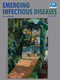
“Many Things Grow in The Garden That Were Never Sown There” [PDF - 2.85 MB - 2 pages]
| EID | Breedlove B. “Many Things Grow in The Garden That Were Never Sown There”. Emerg Infect Dis. 2019;25(11):2162-2163. https://doi.org/10.3201/eid2511.ac2511 |
|---|---|
| AMA | Breedlove B. “Many Things Grow in The Garden That Were Never Sown There”. Emerging Infectious Diseases. 2019;25(11):2162-2163. doi:10.3201/eid2511.ac2511. |
| APA | Breedlove, B. (2019). “Many Things Grow in The Garden That Were Never Sown There”. Emerging Infectious Diseases, 25(11), 2162-2163. https://doi.org/10.3201/eid2511.ac2511. |


