Volume 25, Number 2—February 2019
Synopsis
Atypical Cowpox Virus Infection in Smallpox-Vaccinated Patient, France
Figure 4
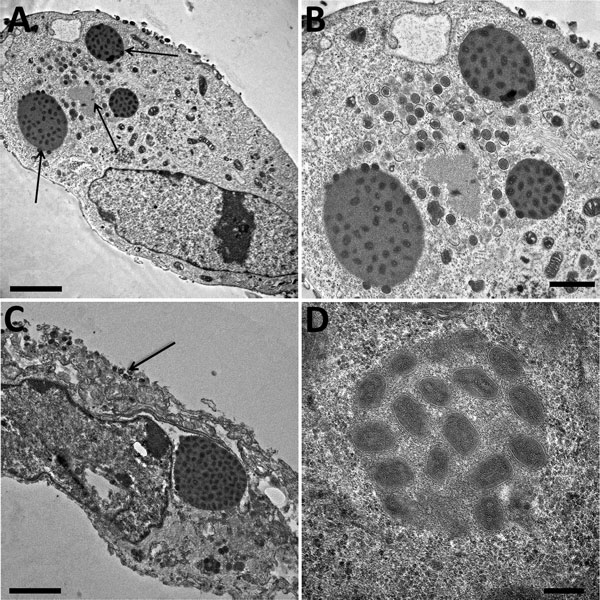
Figure 4. Electron microscopy imaging of cowpox virus France Amiens 2016, obtained from a smallpox-vaccinated patient in France in 2016. A) Ultrathin sections of a Hep2 cell at 32 hours postinfection. The cell harbors, which is undergoing its replicative cycle. Arrows indicate dense inclusion bodies as well as its viral factory containing viral crescents in the cell cytoplasm. Scale bar indicates 2 μm. B) Higher magnification of Hep2 cell in panel A; scale bar indicates 1 μm. C) Ultrathin sections of a Hep2 cell with a typical inclusion of cowpox virus detected near the nucleus. Arrow indicates extracellular-enveloped viruses or cell-associated enveloped particles. Scale bar indicates 2 μm. D) Electron-dense inclusion body containing mature viral particles. Scale bars indicate 200 nm.
1These first authors contributed equally to this article.