Skip directly to site content Skip directly to page options Skip directly to A-Z link Skip directly to A-Z link Skip directly to A-Z link
Volume 25, Number 6—June 2019
Research Letter

Highly Pathogenic Swine Getah Virus in Blue Foxes, Eastern China, 2017

Ning Shi, Li-Xia Li, Rong-Guang Lu, Xi-Jun Yan, and Hao LiuComments to Author 
Author affiliations: Foshan University, Foshan, China (N. Shi, H. Liu); Jilin Wildlife Rescue and Rehabilitation Center, Forestry Department of Jilin Province, Changchun, China (L.-X. Li); Chinese Academy of Agricultural Sciences, Changchun (R.-G. Lu, X.-J. Yan)

Main Article

Figure

Dissected brain and lung of a dead fox, collected in 2017 in Shandong Province, eastern China, and histopathologic examination of samples using hematoxylin and eosin staining. A) Brain, showing congestion in the meninx. B) Histologic view of meninx, showing mild neuronal degeneration and inflammatory cell infiltration in vessels. Original magnification ×100. Box indicates area enlarged in panel C. C) A higher magnification view (original magnification ×400) of lesions in panel B, showing inflamm

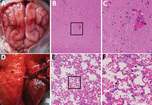
Figure. Dissected brain and lung of a dead fox, collected in 2017 in Shandong Province, eastern China, and histopathologic examination of samples using hematoxylin and eosin staining. A) Brain, showing congestion in the meninx. B) Histologic view of meninx, showing mild neuronal degeneration and inflammatory cell infiltration in vessels. Original magnification ×100. Box indicates area enlarged in panel C. C) A higher magnification view (original magnification ×400) of lesions in panel B, showing inflammatory cell infiltration in a vessel (arrow). D) Lung tissue, showing extensive congestion and hemorrhage. E) Histologic view of lung tissue, showing congestion, hemorrhage, or both, with many erythrocytes in the alveolar space. Original magnification ×100. Box indicates area enlarged in panel F. F) A higher magnification view (original magnification ×400) of tissue lesions in panel E, showing erythrocytes in the alveolar space (arrows).

Main Article

Page created: May 20, 2019
Page updated: May 20, 2019
Page reviewed: May 20, 2019
The conclusions, findings, and opinions expressed by authors contributing to this journal do not necessarily reflect the official position of the U.S. Department of Health and Human Services, the Public Health Service, the Centers for Disease Control and Prevention, or the authors' affiliated institutions. Use of trade names is for identification only and does not imply endorsement by any of the groups named above.
file_external