Volume 25, Number 8—August 2019
Research Letter
Polio-Like Manifestation of Powassan Virus Infection with Anterior Horn Cell Involvement, Canada
Christopher Picheca1, Vignan Yogendrakumar1 , James I. Brooks, Carlos Torres, Elizabeth Pringle, and Jocelyn Zwicker
Figure
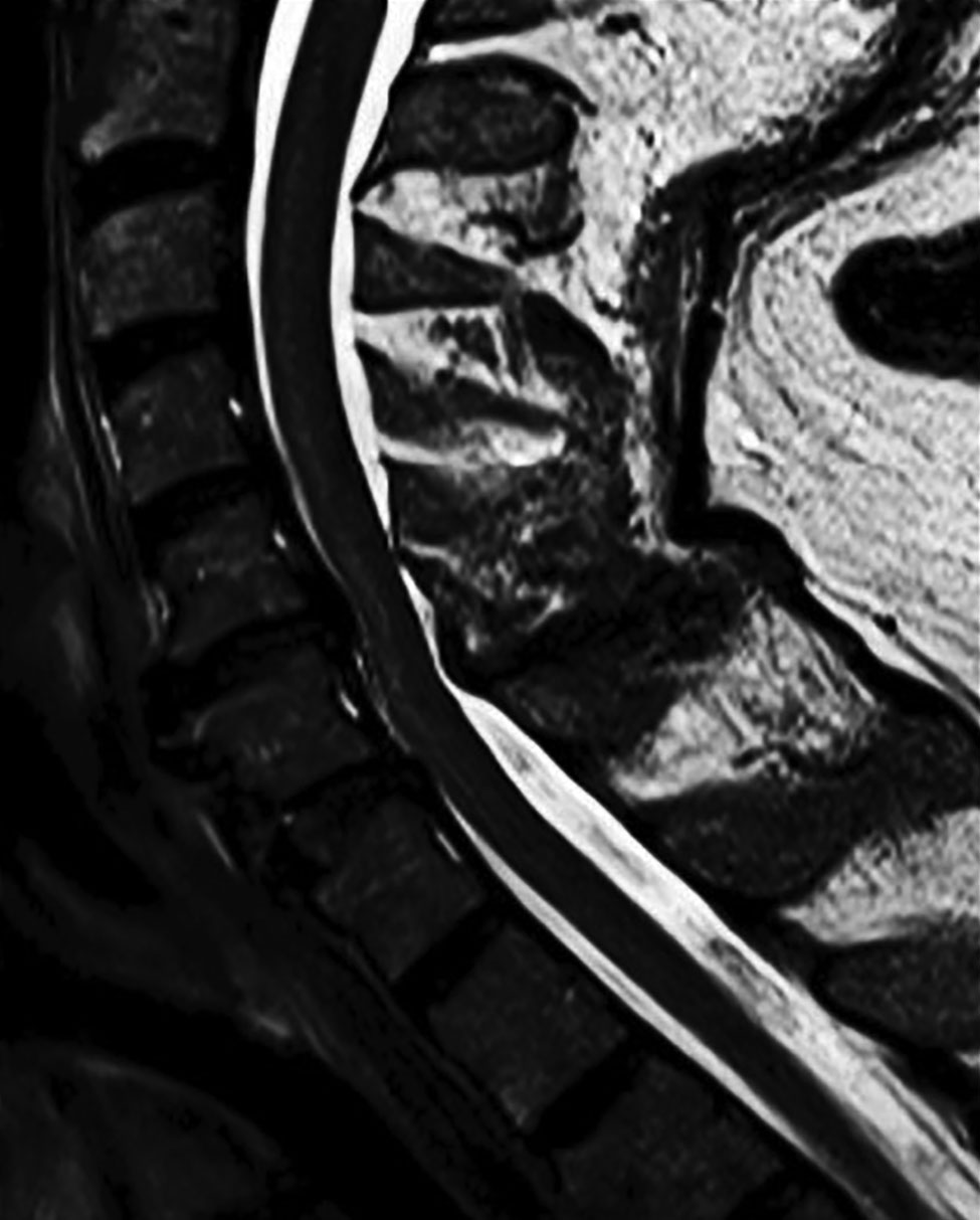
Figure. Sagittal T2-weighted image of cervical spinal cord in a patient with Powassan virus infection, Canada. A longitudinal hyperintensity of the anterior horn is visible from C3 to C6.
1These first authors contributed equally to this article.
Page created: July 17, 2019
Page updated: July 17, 2019
Page reviewed: July 17, 2019
The conclusions, findings, and opinions expressed by authors contributing to this journal do not necessarily reflect the official position of the U.S. Department of Health and Human Services, the Public Health Service, the Centers for Disease Control and Prevention, or the authors' affiliated institutions. Use of trade names is for identification only and does not imply endorsement by any of the groups named above.