Volume 26, Number 10—October 2020
Dispatch
Emerging Sand Fly–Borne Phlebovirus in China
Jing Wang1, Shihong Fu1, Ziqian Xu1, Jingxia Cheng1, Mang Shi, Na Fan, Jingdong Song, Xiaodong Tian, Jianshu Cheng, Shuqing Ni, Ying He, Wenwen Lei, Fan Li, Heng Peng, Bin Wang, Huanyu Wang, Xiaoqing Lu, Yajun Ma , and Guodong Liang
Figure 1
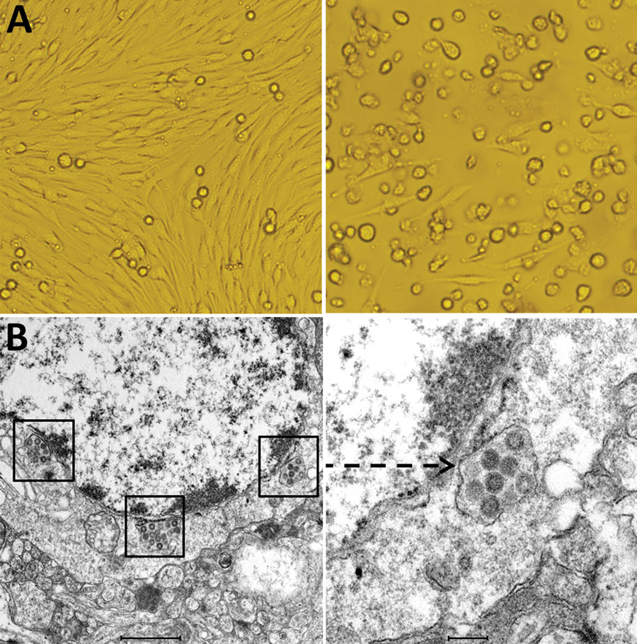
Figure 1. Cytopathogenic effect and electron microscopic morphology of baby hamster kidney 21 (BHK-21) cells infected with phlebovirus, China. A) Left panel shows morphology of BHK-21 cells before inoculation with strain SXWX1813-2; right panel shows morphology 3 days after inoculation (original magnification ×200). BHK-21 cells infected with SXWX1813-2 showed reduced adherence and a large number of rounded and exfoliated cells. B) Left panel shows the viral morphology of SXWX1813–2 on ultrathin slices (scale bar 1μm); right panel shows the enlarged viral particle (indicated by arrow) (scale bar 200nm).
1These authors contributed equally to this article.
Page created: June 30, 2020
Page updated: September 17, 2020
Page reviewed: September 17, 2020
The conclusions, findings, and opinions expressed by authors contributing to this journal do not necessarily reflect the official position of the U.S. Department of Health and Human Services, the Public Health Service, the Centers for Disease Control and Prevention, or the authors' affiliated institutions. Use of trade names is for identification only and does not imply endorsement by any of the groups named above.