Mycoplasma bovis Infections in Free-Ranging Pronghorn, Wyoming, USA
Jennifer L. Malmberg, Donal O’Toole, Terry Creekmore, Erika Peckham, Hally Killion, Madison Vance, Rebecca Ashley, Marguerite Johnson, Christopher Anderson, Marce Vasquez, Douglas Sandidge, Jim Mildenberger, Noah Hull, Dan Bradway, Todd Cornish, Karen B. Register, and Kerry S. Sondgeroth
Author affiliations: Wyoming State Veterinary Laboratory, Laramie, Wyoming, USA (J.L. Malmberg, D. O’Toole, H. Killion, M. Vance, R. Ashley, M. Vasquez, T. Cornish, K.S. Sondgeroth); University of Wyoming Department of Veterinary Sciences, Laramie (J.L. Malmberg, D. O’Toole, M. Johnson, C. Anderson, D. Sandidge, T. Cornish, K.S. Sondgeroth); Wyoming Game and Fish Department, Laramie (T. Creekmore); Wyoming Game and Fish Department, Gillette, Wyoming, USA (E. Peckham); Wyoming Public Health Laboratories, Cheyenne, Wyoming, USA (J. Mildenberger, N. Hull); Washington Animal Disease Diagnostic Laboratory, Pullman, Washington, USA (D. Bradway); US Department of Agriculture National Animal Disease Center, Ames, Iowa, USA (K.B. Register)
Main Article
Figure 4
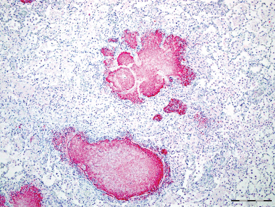
Figure 4. Caseonecrotic lung lesions in free-ranging pronghorn found to be strongly immunopositive for Mycoplasma bovis antigen by immunohistochemical analysis, Wyoming, USA, February–April 2019. Positive staining indicated by fast red coloring has strong intensity and specificity for lesions centered on bronchioles. Scale bar indicates 1 mm.
Main Article
Page created: September 30, 2020
Page updated: November 19, 2020
Page reviewed: November 19, 2020
The conclusions, findings, and opinions expressed by authors contributing to this journal do not necessarily reflect the official position of the U.S. Department of Health and Human Services, the Public Health Service, the Centers for Disease Control and Prevention, or the authors' affiliated institutions. Use of trade names is for identification only and does not imply endorsement by any of the groups named above.