Skip directly to site content Skip directly to page options Skip directly to A-Z link Skip directly to A-Z link Skip directly to A-Z link
Volume 26, Number 12—December 2020
Dispatch

Hypervirulent Klebsiella pneumoniae as Unexpected Cause of Fatal Outbreak in Captive Marmosets, Brazil

Juliana Mariotti Guerra, Natália Coelho Couto de A. Fernandes, Alessandra Loureiro Morales dos Santos, Joana de Souza Pereira Barrel, Bruno Simões Sergio Petri, Liliane Milanelo, Monique Ribeiro Tiba-Casas, Alcina Maria Liserre, Cláudia Regina Gonçalves, Cláudio Tavares Sacchi, José Luiz Catão-Dias, and Carlos Henrique CamargoComments to Author 
Author affiliations: Instituto Adolfo Lutz, São Paulo, Brazil (J.M. Guerra, N.C.C.A. Fernandes, A.L.M. dos Santos, J.S.P. Barrel, M.R. Tiba-Casas, A.M. Liserre, C.R Gonçalves., C.T Sacchi., C.H. Camargo); Universidade de São Paulo, São Paulo (N.C.C.A. Fernandes, A.L.M. dos Santos, J.L. Catão-Dias); Parque Ecológico do Tietê, São Paulo (B.S.S. Petri, L. Milanelo)

Main Article

Figure 1

Microscopic findings of histological and histochemical examination of tissue samples from captive marmosets in investigation of a fatal epizootic caused by highly virulent Klebsiella pneumoniae sequence type 86 strain P04 in Brazil, 2019. A) Spleen shows necrosis in germinal centers, suppurative splenitis, and hemorrhage (inset: necrosis in germinal center). Hematoxylin and eosin stain (H&E); original magnification ×4. B) Brain (meninges) shows bacterial rods inside vascular lumen (arrow). H&E stain; original magnification ×40. C) Lung shows sinterstitial pneumonia (H&E stain; original magnification ×4) and alveolar hemorrhage (inset; H&E stain; original magnification ×10). D–F) Liver samples. D) Numerous intravascular bacilli (arrow). H&E stain; original magnification ×100. E) Hepatocellular necrosis (arrowheads) associated with numerous bacterial rods (arrow) and neutrophils in the sinusoids. H&E stain; original magnification ×40. F) Sinusoids filled with gram-negative bacterial structures (arrow) and neutrophils. Gram stain; original magnification ×1,000.

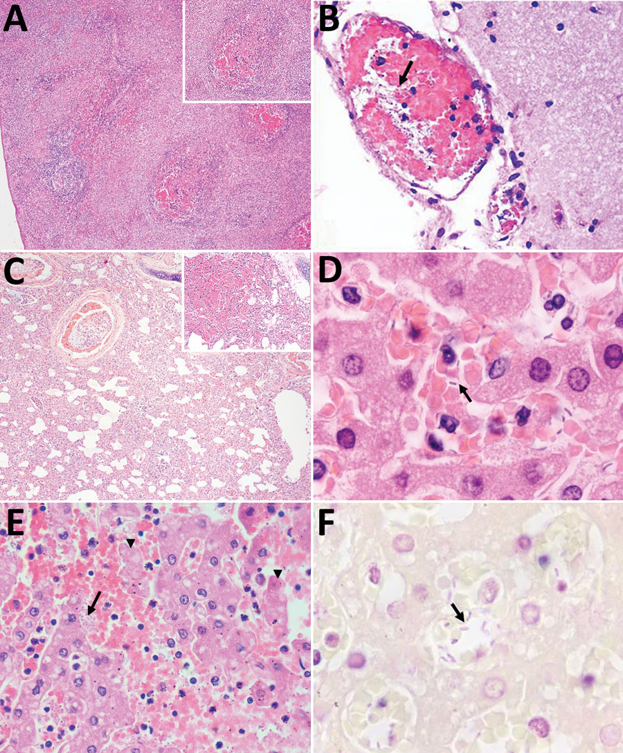
Figure 1. Microscopic findings of histological and histochemical examination of tissue samples from captive marmosets in investigation of a fatal epizootic caused by highly virulent Klebsiella pneumoniae sequence type 86 strain P04 in Brazil, 2019. A) Spleen shows necrosis in germinal centers, suppurative splenitis, and hemorrhage (inset: necrosis in germinal center). Hematoxylin and eosin stain (H&E); original magnification ×4. B) Brain (meninges) shows bacterial rods inside vascular lumen (arrow). H&E stain; original magnification ×40. C) Lung shows sinterstitial pneumonia (H&E stain; original magnification ×4) and alveolar hemorrhage (inset; H&E stain; original magnification ×10). D–F) Liver samples. D) Numerous intravascular bacilli (arrow). H&E stain; original magnification ×100. E) Hepatocellular necrosis (arrowheads) associated with numerous bacterial rods (arrow) and neutrophils in the sinusoids. H&E stain; original magnification ×40. F) Sinusoids filled with gram-negative bacterial structures (arrow) and neutrophils. Gram stain; original magnification ×1,000.

Main Article

Page created: October 13, 2020
Page updated: November 19, 2020
Page reviewed: November 19, 2020
The conclusions, findings, and opinions expressed by authors contributing to this journal do not necessarily reflect the official position of the U.S. Department of Health and Human Services, the Public Health Service, the Centers for Disease Control and Prevention, or the authors' affiliated institutions. Use of trade names is for identification only and does not imply endorsement by any of the groups named above.
file_external