Skip directly to site content Skip directly to page options Skip directly to A-Z link Skip directly to A-Z link Skip directly to A-Z link
Volume 26, Number 12—December 2020
Dispatch

Shedding of Marburg Virus in Naturally Infected Egyptian Rousette Bats, South Africa, 2017

Janusz T. PawęskaComments to Author , Nadia Storm, Wanda Markotter, Nicholas Di Paola, Michael R. Wiley, Gustavo Palacios, and Petrus Jansen van Vuren
Author affiliations: National Institute for Communicable Diseases of the National Health Laboratory Service, Johannesburg, South Africa (J.T. Pawęska, N. Storm, P. Jansen van Vuren); Boston University, Boston, Massachusetts, USA (N. Storm); University of Pretoria, Pretoria, South Africa (J.T. Pawęska, W. Markotter); University of Nebraska Medical Center, Omaha, Nebraska, USA (N. Di Paola, M.R. Wiley, G. Palacios); US Army Medical Research Institute of Infectious Diseases, Frederick, Maryland, USA (N. Di Paola, M.R. Wiley, G. Palacios); Australian Centre for Disease Preparedness, Commonwealth Scientific and Industrial Research Organisation–Health and Biosecurity, Geelong, Victoria, Australia (P. Jansen van Vuren)

Main Article

Figure 1

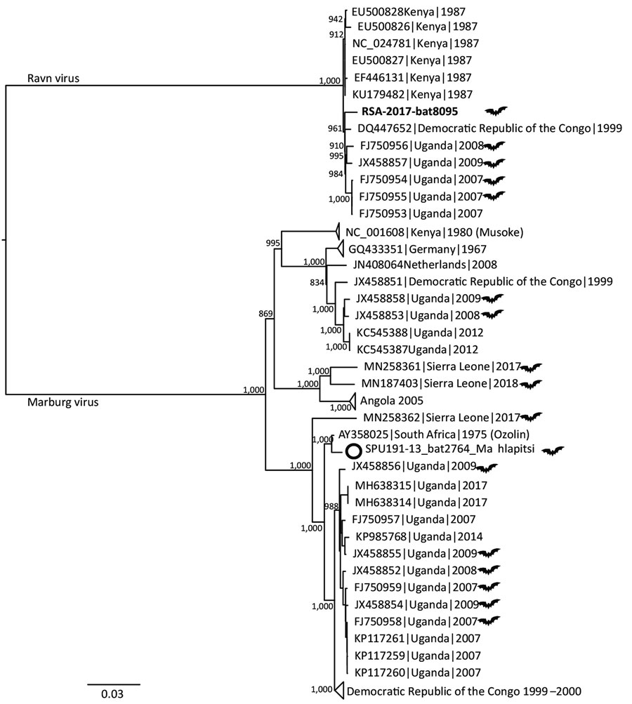
Midpoint-rooted, maximum-likelihood phylogeny of complete and near-complete MARV and RAVV genomes. Phylogenetic tree shows evolutionary relationships of marburgvirus detected in a rectal swab sample from a subadult Egyptian rousette female bat (Rousettus aegyptiacus) in Matlapisi Cave, Limpopo Province, South Africa, 2017 (black filled circle; GenBank accession no. MT321489), and reference viruses, including the SPU191-13 bat 2764 Mahlapitsi strain (white circle; GenBank accession no. MG725616), detected in the same cave in July 2013. Complete and near-complete genome sequences from GenBank (accession numbers indicated) were aligned with the partial MARV sequence obtained from RSA-8095bat using MUSCLE version 3.8.31 (https://www.drive5.com/muscle), and RAxML version 8.2.10 (https://cme.h-its.org/exelixis/web/software/raxml/index.html) was used to infer the best-scoring maximum-likelihood tree after 1,000 bootstrap replicates. Node values indicate the bootstrap support values. Genomes isolated from bats are shown using a bat symbol. Scale bar indicates nucleotide substitutions per site. MARV, Marburg virus; RAVV, Ravn virus.

Figure 1. Midpoint-rooted, maximum-likelihood phylogeny of complete and near-complete MARV and RAVV genomes. Phylogenetic tree shows evolutionary relationships of marburgvirus detected in a rectal swab sample from a subadult Egyptian rousette female bat (Rousettus aegyptiacus) in Matlapisi Cave, Limpopo Province, South Africa, 2017 (black filled circle; GenBank accession no. MT321489), and reference viruses, including the SPU191-13 bat 2764 Mahlapitsi strain (white circle; GenBank accession no. MG725616), detected in the same cave in July 2013. Complete and near-complete genome sequences from GenBank (accession numbers indicated) were aligned with the partial MARV sequence obtained from RSA-8095bat using MUSCLE version 3.8.31 (https://www.drive5.com/muscle), and RAxML version 8.2.10 (https://cme.h-its.org/exelixis/web/software/raxml/index.html) was used to infer the best-scoring maximum-likelihood tree after 1,000 bootstrap replicates. Node values indicate the bootstrap support values. Genomes isolated from bats are shown using a bat symbol. Scale bar indicates nucleotide substitutions per site. MARV, Marburg virus; RAVV, Ravn virus.

Main Article

Page created: August 12, 2020
Page updated: November 19, 2020
Page reviewed: November 19, 2020
The conclusions, findings, and opinions expressed by authors contributing to this journal do not necessarily reflect the official position of the U.S. Department of Health and Human Services, the Public Health Service, the Centers for Disease Control and Prevention, or the authors' affiliated institutions. Use of trade names is for identification only and does not imply endorsement by any of the groups named above.
file_external