Volume 26, Number 12—December 2020
Research Letter
Transmission Electron Microscopy Confirmation of Orientia tsutsugamushi in Human Bile
Figure

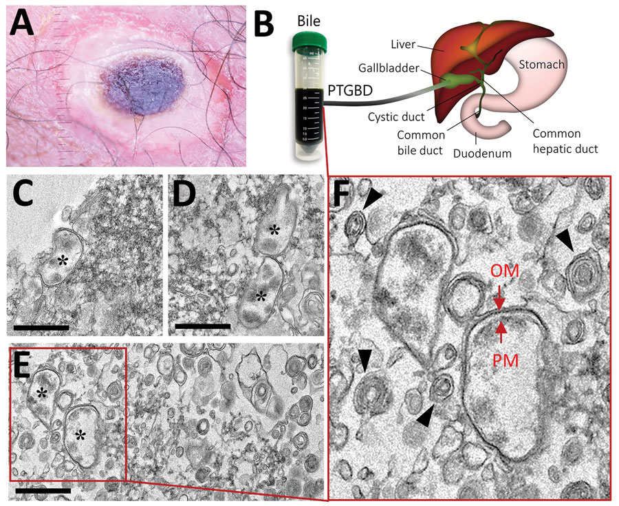
Figure. Findings from a 68-year-old woman with scrub typhus, South Korea, 2019. A) Eschar in the right inguinal area. B) Human bile collected through percutaneous transhepatic gallbladder drainage in the gallbladder of a patient affected with scrub typhus. C–F) Transmission electron microscopy images of Orientia tsutsugamushi in the bile. Bacteria (black asterisks); outer membrane (OM) and plasma membrane (PM) (red arrows); multilamellar body (black arrowheads); Scale bars indicate 1 μm.
1These authors contributed equally to this article.
2These authors were co-principal investigators.
Page created: October 16, 2020
Page updated: November 19, 2020
Page reviewed: November 19, 2020
The conclusions, findings, and opinions expressed by authors contributing to this journal do not necessarily reflect the official position of the U.S. Department of Health and Human Services, the Public Health Service, the Centers for Disease Control and Prevention, or the authors' affiliated institutions. Use of trade names is for identification only and does not imply endorsement by any of the groups named above.