Volume 26, Number 3—March 2020
Dispatch
Progressive Vaccinia Acquired through Zoonotic Transmission in a Patient with HIV/AIDS, Colombia
Katherine Laiton-Donato1 , Paola Ávila-Robayo1, Andrés Páez-Martinez, Paula Benjumea-Nieto, José A. Usme-Ciro, Nicole Pinzón-Nariño, Ivan Giraldo, Diego Torres-Castellanos, Yoshinori Nakazawa, Nishi Patel, Kimberly Wilkins, Yu Li, Whitni Davidson, Jillybeth Burgado, Panayampalli Subbian Satheshkumar, Ashley Styczynski, Matthew R. Mauldin, Martha Gracia-Romero, and Brett W. Petersen
Figure 1
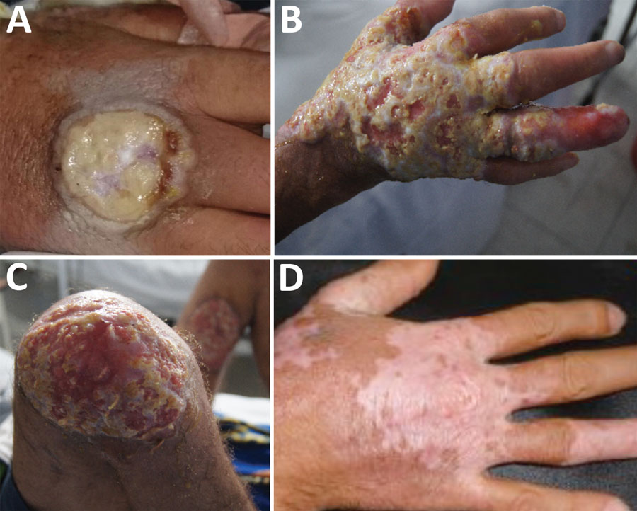
Figure 1. Clinical progression of vaccinia virus infection in a patient with HIV/AIDS, Colombia. A) On December 9, 2014, the patient was referred to the Hospital Universitario Mayor Méderi because of a suppurative ulcer with sharply raised, defined edges on his right hand. B, C) In March 2015, lesions increased in size and disseminated over his face and extremities. D) In July 2015, most lesions completely healed, with mild scarring and depigmentation.
1These authors contributed equally to this article.
Page created: February 20, 2020
Page updated: February 20, 2020
Page reviewed: February 20, 2020
The conclusions, findings, and opinions expressed by authors contributing to this journal do not necessarily reflect the official position of the U.S. Department of Health and Human Services, the Public Health Service, the Centers for Disease Control and Prevention, or the authors' affiliated institutions. Use of trade names is for identification only and does not imply endorsement by any of the groups named above.