Volume 26, Number 5—May 2020
Research Letter
Diplorickettsia Bacteria in an Ixodes scapularis Tick, Vermont, USA
Cite This Article
Citation for Media
Abstract
An unexpected Diplorickettsia species closely related to the tickborne pathogen D. massieliensis was found in the microbiome of an Ixodes scapularis tick in Vermont, USA. This evidence of Diplorickettsia in North American ticks suggests a need for disease surveillance using molecular screening of ticks and serologic studies of humans.
The blacklegged tick, Ixodes scapularis, is a generalist arthropod ectoparasite that serves as a vector for an array of common human pathogens; novel disease-causing microbes have been discovered consistently in the tick for the past several decades. Tickborne bacterial infections causing illnesses such as anaplasmosis, Borrelia miyamotoi disease, and ehrlichiosis have all emerged in the United States in recent years (1–3), and it has been estimated that as many as half of all tickborne illnesses are caused by unknown pathogens (4).
We used 16S rRNA sequencing to survey for bacterial pathogens in I. scapularis ticks in western Vermont, USA. We collected ticks by drag sampling along 100 m transects using a 1 m2 square of white denim. We collected ticks from 6 deciduous forest sites in Addison and Chittenden counties, Vermont (Appendix Table 1), during May–July 2015. We extracted DNA from ticks using a phenol-chloroform extraction (5). In total, we extracted DNA from 97 ticks, 20 of which we selected based on DNA quality and quantity for 16S rDNA sequencing at Hudson Alpha Genomic Services Laboratory (Huntsville, AL, USA). We PCR amplified the V3 and V4 regions of the 16S rRNA gene from these ticks and 1 blank using 341F and 875R primers (6) and sequenced them on a MiSeq platform (Illumina, https://www.illumina.com), yielding a total of 15,302,568 reads. We used the DADA2 R package to identify amplicon sequence variants (ASVs) and assign taxonomy (7) (Appendix Figure 1). We used default settings in the DADA2 pipeline; however, we estimated error rates using the first 10 billion base pairs.
Figure
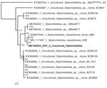
Figure. Neighbor-joining phylogenetic tree of a MAFFT alignment (https://mafft.cbrc.jp/alignment/server) of the V3–V4 region of the Diplorickettsia 16S rRNA gene, including the novel amplicon sequence variant identified in Vermont, USA (bold). A...
In a single adult male tick, an ASV assigned to the genus Diplorickettsia comprised 82% of the microbiome sequencing reads. The genus Diplorickettsia was originally defined by the species D. massiliensis, discovered in I. ricinus ticks in Europe (8). The ASV we identified shared 425 of 427 nt in the sequenced V3–V4 region of the 16S rRNA gene with the reference sequence of D. massiliensis strain 20B (GenBank accession no. NR_117407.1) and was more closely related to this strain than any other previously sequenced Diplorickettsia in the National Center for Biotechnology Information (NCBI) nucleotide database (Figure). This tick was collected at the Sunny Hollow Colchester site (coordinates 44.518353°, −73.17112°) (Appendix Table 1).
D. massiliensis has been identified as a possible human pathogen; of patients in a hospital in France, 3 were found seropositive, and 1 found positive by quantitative PCR for the D. massiliensis rpoB gene (9). Our findings represent evidence of a Diplorickettsia bacteria in ticks in North America.
To confirm the presence of Diplorickittsia in the positive tick, we designed PCR primers to regions of the D. massiliensis parC and ftsY genes (Appendix Table 2) using primer-BLAST (https://www.ncbi.nlm.nih.gov/tools/primer-blast). We aligned primers against the NCBI nr database to ensure specific binding to Diplorickettsia. We also used DNA from a Diplorickettsia-negative tick (as determined by 16S sequencing) as PCR template to serve as a negative control. Successful amplification of regions of both genes confirmed the presence of Diplorickettsia DNA in the positive tick (Appendix Figure 2).
We Sanger sequenced amplicons from these PCR tests. We combined forward and reverse reads and trimmed them using the PEAR utility (https://cme.h-its.org/exelixis/web/software/pear). Each sequence showed high identity with previously sequenced D. massiliensis reference sequences via gapped alignment (247/252 bp parC, 298/310 bp ftsY). These results further suggest a close relationship between Diplorickettsia species we identified and D. massiliensis, but a lack of reference sequences for these genes from other species of Diplorickettsia makes it impossible to definitively assign this uncultured specimen to a particular species.
The high sequence similarity between the Diplorickettsia we identified and the previously identified pathogenic variety suggests the need for further study of the pathogenicity of this variant. Many genera of tickborne bacteria contain both pathogenic and nonpathogenic strains, and genetic similarity alone cannot confirm pathogenicity. Future work is needed to isolate this strain of Diplorickettsia and determine its ability to infect mammalian hosts and its transmissibility via tick bite. Experiments to test its ability to induce febrile illness in mammals would also help determine if Diplorickettsia spp. could cause a clinically significant infection in humans. Furthermore, serologic studies of patients with suspected tickborne diseases in the area surrounding the collection site are necessary to determine if this bacterium has infected persons in Vermont.
In addition, our findings suggest the need for further study of the prevalence of Diplorickettsia in North America ticks. We have developed PCR primers (Appendix Table 2) to facilitate future study of this bacterium and have demonstrated via sequencing that these primers accurately amplify their target Diplorickettsia genes. We have deposited the partial Diplorickettsia 16S rRNA, ftsY, and parC genes sequenced in this study into the NCBI GenBank database (accession nos. MN192053, MN640996, MN640997). The raw sequencing reads are available through the NCBI sequence read archive (accession no. PRJNA557440).
Mr. Merenstein is a computational analyst at Boston University School of Medicine, Boston, Massachusetts. His primary interests include microbial ecology and host-associated microbes.
Acknowledgment
Research reported in this publication was supported by an Institutional Development Award (IDeA) from the National Institute of General Medical Sciences of the National Institutes of Health under grant no. P20GM103449. Additional funding was provided by the Middlebury College Biology Department and the Albert D. Mead Professorship at Middlebury College.
References
- Buller RS, Arens M, Hmiel SP, Paddock CD, Sumner JW, Rikhisa Y, et al. Ehrlichia ewingii, a newly recognized agent of human ehrlichiosis. N Engl J Med. 1999;341:148–55. DOIPubMedGoogle Scholar
- Dumler JS, Choi K-S, Garcia-Garcia JC, Barat NS, Scorpio DG, Garyu JW, et al. Human granulocytic anaplasmosis and Anaplasma phagocytophilum. Emerg Infect Dis. 2005;11:1828–34. DOIPubMedGoogle Scholar
- Telford SR III, Goethert HK, Molloy PJ, Berardi VP, Chowdri HR, Gugliotta JL, et al. Borrelia miyamotoi disease: neither Lyme disease nor relapsing fever. Clin Lab Med. 2015;35:867–82. DOIPubMedGoogle Scholar
- Feder HM Jr, Johnson BJB, O’Connell S, Shapiro ED, Steere AC, Wormser GP; Ad Hoc International Lyme Disease Group. A critical appraisal of “chronic Lyme disease”. N Engl J Med. 2007;357:1422–30. DOIPubMedGoogle Scholar
- Van Treuren W, Ponnusamy L, Brinkerhoff RJ, Gonzalez A, Parobek CM, Juliano JJ, et al. Variation in the microbiota of Ixodes ticks with regard to geography, species, and sex. Appl Environ Microbiol. 2015;81:6200–9. DOIPubMedGoogle Scholar
- Klindworth A, Pruesse E, Schweer T, Peplies J, Quast C, Horn M, et al. Evaluation of general 16S ribosomal RNA gene PCR primers for classical and next-generation sequencing-based diversity studies. Nucleic Acids Res. 2013;41:
e1 . DOIPubMedGoogle Scholar - Callahan BJ, McMurdie PJ, Rosen MJ, Han AW, Johnson AJA, Holmes SP. DADA2: High-resolution sample inference from Illumina amplicon data. Nat Methods. 2016;13:581–3. DOIPubMedGoogle Scholar
- Mediannikov O, Sekeyová Z, Birg M-L, Raoult D. A novel obligate intracellular gamma-proteobacterium associated with ixodid ticks, Diplorickettsia massiliensis, Gen. Nov., Sp. Nov. PLoS One. 2010;5:
e11478 . DOIPubMedGoogle Scholar - Subramanian G, Mediannikov O, Angelakis E, Socolovschi C, Kaplanski G, Martzolff L, et al. Diplorickettsia massiliensis as a human pathogen. Eur J Clin Microbiol Infect Dis. 2012;31:365–9. DOIPubMedGoogle Scholar
Figure
Cite This ArticleOriginal Publication Date: April 06, 2020
Table of Contents – Volume 26, Number 5—May 2020
| EID Search Options |
|---|
|
|
|
|
|
|
Please use the form below to submit correspondence to the authors or contact them at the following address:
David Allen, Department of Biology, Middlebury College, McCardell Bicentennial Hall 372, Middlebury, VT 05753, USA
Top