Volume 26, Number 6—June 2020
Dispatch
No Adaptation of the Prion Strain in a Heterozygous Case of Variant Creutzfeldt-Jakob Disease
Figure 2
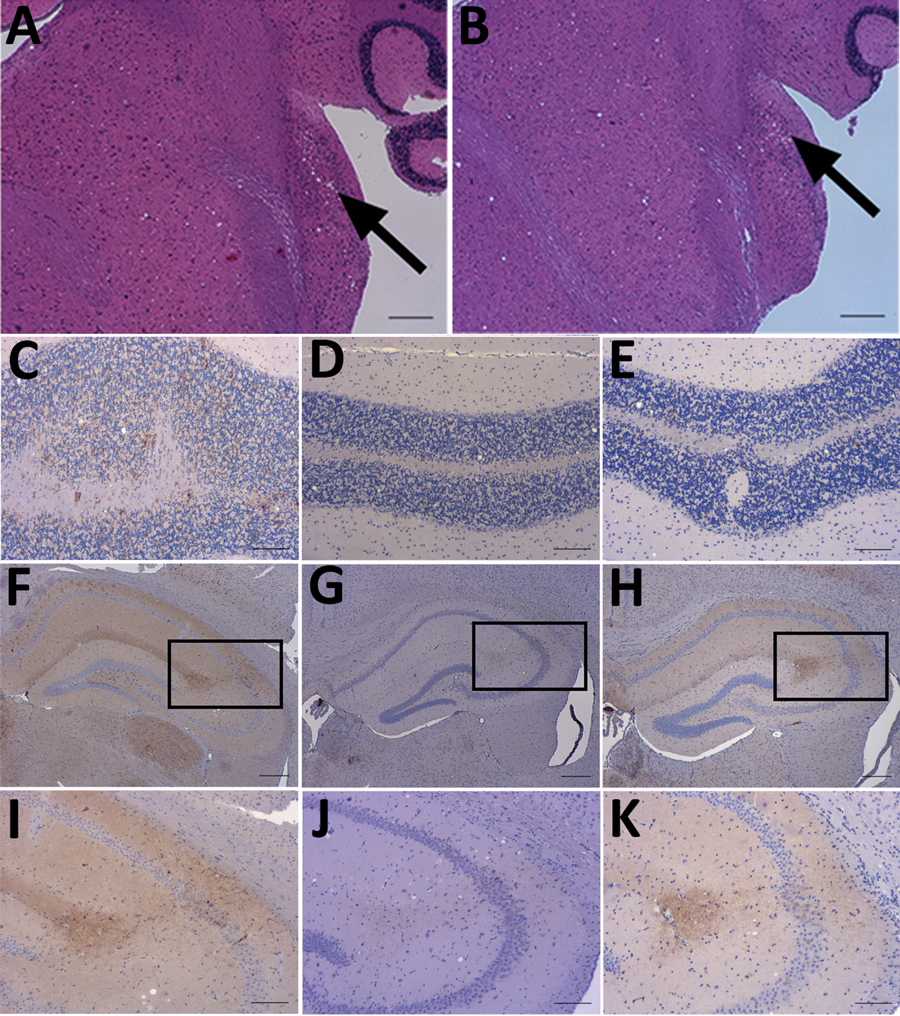
Figure 2. Neuropathology of RIII mice inoculated with material from a clinical case of vCJD in a prion protein gene codon 129MV individual, a typical 129MM case of vCJD, and BSE. A, B) Haemotoxylin and Eosin staining of transmissible spongiform encephalopathy vacuolation in the cochlear nucleus of mice inoculated with material from a clinical 129MV case (arrows). C–E) Abnormal PrP deposition in the cerebellum of mice inoculated with C) clinical 129MV case, D) typical 129MM case, and E) BSE. F–H) Abnormal PrP deposition in the hippocampus of mice inoculated with samples of F) clinical 129MV case, G) typical 129MM case, and H) BSE. I–K) Abnormal PrP deposition in the CA2 region of the hippocampus; I) inset from panel F; J) inset from panel G; K) inset from panel H. Monoclonal antibody: 6H4. Scale bars: A–B, F–H: 200 µm. C–E, I–K: 100 µm. BSE, bovine spongiform encephalopathy; MM, methionine homozygous; MV, methionine/valine; PrP, prion protein; vCJD, variant Creutzfeldt-Jakob disease.