Volume 26, Number 6—June 2020
Research
Severe Acute Respiratory Syndrome Coronavirus 2 from Patient with Coronavirus Disease, United States
Jennifer Harcourt1, Azaibi Tamin1, Xiaoyan Lu, Shifaq Kamili, Senthil K. Sakthivel, Janna Murray, Krista Queen, Ying Tao, Clinton R. Paden, Jing Zhang, Yan Li, Anna Uehara, Haibin Wang, Cynthia Goldsmith, Hannah A. Bullock, Lijuan Wang, Brett Whitaker, Brian Lynch, Rashi Gautam, Craig Schindewolf, Kumari G. Lokugamage, Dionna Scharton, Jessica A. Plante, Divya Mirchandani, Steven G. Widen, Krishna Narayanan, Shinji Makino, Thomas G. Ksiazek, Kenneth S. Plante, Scott C. Weaver, Stephen Lindstrom, Suxiang Tong, Vineet D. Menachery2, and Natalie J. Thornburg2
Figure 1
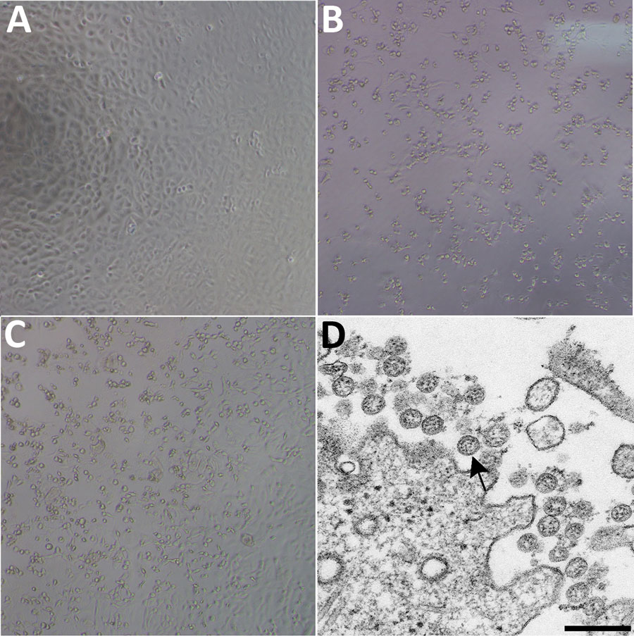
Figure 1. Cytopathic effect caused by severe acute respiratory syndrome coronavirus 2 from patient with coronavirus disease, United States, 2020. A–C) Phase-contrast microscopy of Vero cell monolayers at 3 days postinoculation: A) Mock, B) nasopharyngeal specimen, C) oropharyngeal specimen. Original magnifications ×10). D) Electron microscopy of virus isolate showing extracellular spherical particles with cross-sections through the nucleocapsids (black dots). Arrow indicates a coronavirus virion budding from a cell. Scale bar indicates 200 nm.
1These authors contributed equally to this article.
2These senior authors contributed equally to this article.
Page created: May 18, 2020
Page updated: May 18, 2020
Page reviewed: May 18, 2020
The conclusions, findings, and opinions expressed by authors contributing to this journal do not necessarily reflect the official position of the U.S. Department of Health and Human Services, the Public Health Service, the Centers for Disease Control and Prevention, or the authors' affiliated institutions. Use of trade names is for identification only and does not imply endorsement by any of the groups named above.