Skip directly to site content Skip directly to page options Skip directly to A-Z link Skip directly to A-Z link Skip directly to A-Z link
Volume 26, Number 8—August 2020
Dispatch

Spread of Multidrug-Resistant Bacteria by Moth Flies from Hospital Waste Water System

Thomas Rupprecht1, Annette Moter, Alexandra Wiessener, Joerg Reutershan, Klaus Lang-Schwarz, Michael Vieth, Christian Rupprecht, Ruediger Wagner, and Thomas Bollinger1Comments to Author 
Author affiliations: Klinikum Bayreuth, Bayreuth, Germany (T. Rupprecht, J. Reutershan, K. Lang-Schwarz, M. Vieth, T. Bollinger); Friedrich-Alexander University, Erlangen, Germany (T. Rupprecht, M. Vieth); Charité-Universitätsmedizin, Berlin, Germany (A. Moter, A. Wiessener); Eberhard Karls University, Tübingen, Germany (J. Reutershan); University of Oxford, Oxford, UK (C. Rupprecht); Universität Kassel, Kassel, Germany (R. Wagner); University Hospital Schleswig-Holstein, Lübeck, Germany (T. Bollinger)

Main Article

Figure 2

Fluorescence in situ hybridization (FISH) from a longitudinal section of a leg of a Clogmia albipunctata moth fly from a hospital in Germany. The fly was caught in the hospital, embedded, and stained (Appendix, https://wwwnc.cdc.gov/EID/article/26/8/19-0750-App1.pdf). A) Overview showing an overlay of the fluorescent images with a phase contrast to visualize the limbs of the legs. B) Higher magnification of the inset from panel A shows the anatomy of the tarsus and claws with an adjacent biofilm

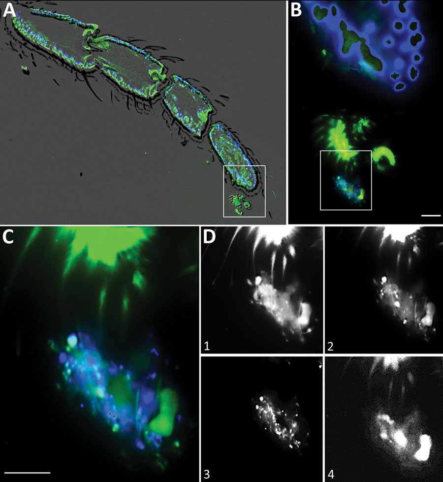
Figure 2. Fluorescence in situ hybridization (FISH) from a longitudinal section of a leg of a Clogmia albipunctata moth fly from a hospital in Germany. The fly was caught in the hospital, embedded, and stained (Appendix, https://wwwnc.cdc.gov/EID/article/26/8/19-0750-App1.pdf). A) Overview showing an overlay of the fluorescent images with a phase contrast to visualize the limbs of the legs. B) Higher magnification of the inset from panel A shows the anatomy of the tarsus and claws with an adjacent biofilm, which is stained by the bacterial probe (green) and DAPI (blue). C) Higher magnification of the inset from panel B shows the biofilm. Blue represents DAPI staining of DNA; bacteria were stained green with pan-bacterial FISH probe EUB338-FITC, Enterobacterales stained orange with an Escherichia coli–specific FISH probe (data not shown), and NONEUB (nonsense EUB) probe labeled with Cy5 was used to exclude unspecific probe binding. D) Overlay of the DAPI and fluorescein isothiocyanate channel shows the biofilm with different bacterial morphotypes. Different planes of the z-stack in the green channel (pan-bacterial probe) of the identical microscopic field depicts the different claws embracing the biofilm (D1 and D2). D3 shows the DAPI filter-set only with the DNA of the bacteria, whereas D4 shows the autofluorescence in the Cy5 filter-set NONEUB probe.

Main Article

1These authors were co–principal investigators.

Page created: April 29, 2020
Page updated: July 18, 2020
Page reviewed: July 18, 2020
The conclusions, findings, and opinions expressed by authors contributing to this journal do not necessarily reflect the official position of the U.S. Department of Health and Human Services, the Public Health Service, the Centers for Disease Control and Prevention, or the authors' affiliated institutions. Use of trade names is for identification only and does not imply endorsement by any of the groups named above.
file_external