Volume 26, Number 8—August 2020
Dispatch
In Vivo Observation of Trombiculosis with Fluorescence–Advanced Videodermatoscopy
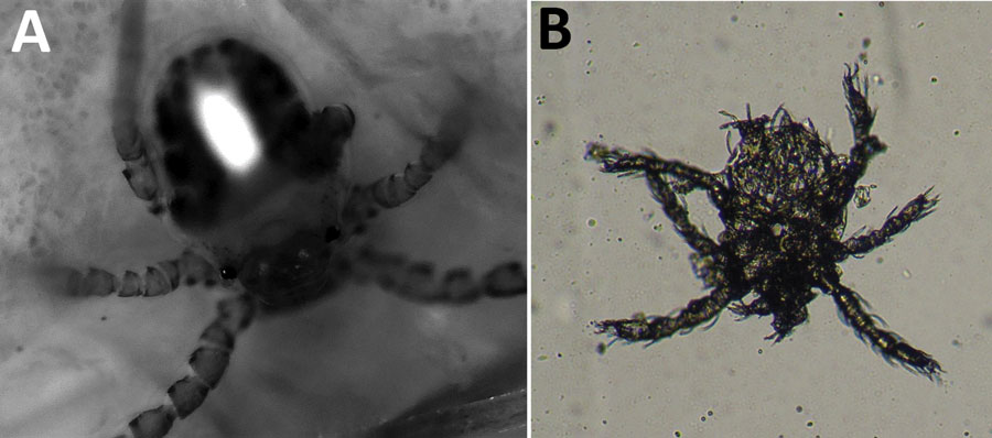
Figure 2

Figure 2. Images of trombiculid mites from infestation of a 48-year-old woman, Italy, April 2019. A) Fluorescence–advanced videodermatoscopy image shows oval body with 3 pairs of legs. Original magnification ×500. B) Optical microscope examination of 1 larva detected from a superficial skin scraping. Original magnification ×100.
1These first authors contributed equally to this article.
2These senior authors contributed equally to this article.
Page created: May 11, 2020
Page updated: July 18, 2020
Page reviewed: July 18, 2020
The conclusions, findings, and opinions expressed by authors contributing to this journal do not necessarily reflect the official position of the U.S. Department of Health and Human Services, the Public Health Service, the Centers for Disease Control and Prevention, or the authors' affiliated institutions. Use of trade names is for identification only and does not imply endorsement by any of the groups named above.