Fatal Measles Inclusion-Body Encephalitis in Adult with Untreated AIDS, France
Christophe Rodriguez, Meriadeg Ar Gouilh, Nicolas Weiss, Sébastian Stroer, Karima Mokhtari, Danielle Seilhean, Bertrand Mathon, Vanessa Demontant, Melissa N’Debi, Guillaume Gricourt, Paul-Louis Woerther, Jean-Michel Pawlotsky, Karl Stefic, Julien Marlet, Pierre-François Dequin, Antoine Guillon, Valérie Pourcher, David Boutolleau, Astrid Vabret, and Sonia Burrel

Author affiliations: Henri Mondor Hospital, Assistance Publique des Hôpitaux de Paris, University of Paris-Est, Créteil, France (C. Rodriguez, V. Demontant, M. N’Debi, G. Gricourt, P.-L. Woerther, J.-M. Pawlotsky); National Reference Laboratory for Measles, Mumps, and Rubella, University Hospital of Caen, Normandie Université, Caen, France (M. Ar Gouilh, A. Vabret); Pitié-Salpêtrière Hospital, Assistance Publique des Hôpitaux de Paris, Sorbonne-Université, Paris, France (N. Weiss, S. Stroer, K. Mokhtari, D. Seilhean, B. Mathon, V. Pourcher, D. Boutolleau, S. Burrel); National Reference Center for Herpesviruses, Paris (D. Boutolleau, S. Burrel); University Hospital of Tours, University of Tours, Tours, France (K. Stefic, J. Marlet, P.-F. Dequin, A. Guillon); National Reference Center for HIV, Tours (K. Stefic, J. Marlet)
Main Article
Figure 1
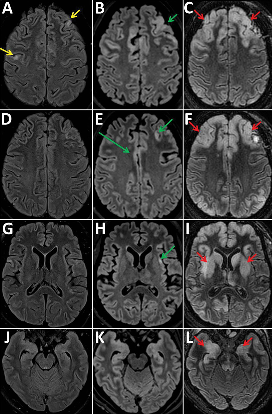
Figure 1. Fluid-attenuated inversion recovery images of magnetic resonance examinations of the brain in a 28-year-old woman from Romania with untreated AIDS and measles inclusion-body encephalitis. Images were taken 1 week (A, D, G, J), 2 weeks (B, E, H, K), and 5 weeks (C, F, I, L) after hospital admission, at the same brain levels. The first examination shows focal cortical hyperintensities (yellow arrows) in the left and right frontal cortex. After 2 weeks, these cortical hyperintensities have widened and are spreading to the cingulum and the insula (green arrows). At 5 weeks, cortical hyperintensities involve a larger part of the neocortex, but also spread to the basal ganglia, amygdala (red arrows), and hippocampus (the hippocampus changes may also be induced by status epilepticus) and to the posterior areas of the pons.
Main Article
Page created: July 02, 2020
Page updated: August 19, 2020
Page reviewed: August 19, 2020
The conclusions, findings, and opinions expressed by authors contributing to this journal do not necessarily reflect the official position of the U.S. Department of Health and Human Services, the Public Health Service, the Centers for Disease Control and Prevention, or the authors' affiliated institutions. Use of trade names is for identification only and does not imply endorsement by any of the groups named above.