Volume 26, Number 9—September 2020
Research Letter
Acute Cerebral Stroke with Multiple Infarctions and COVID-19, France, 2020
Figure
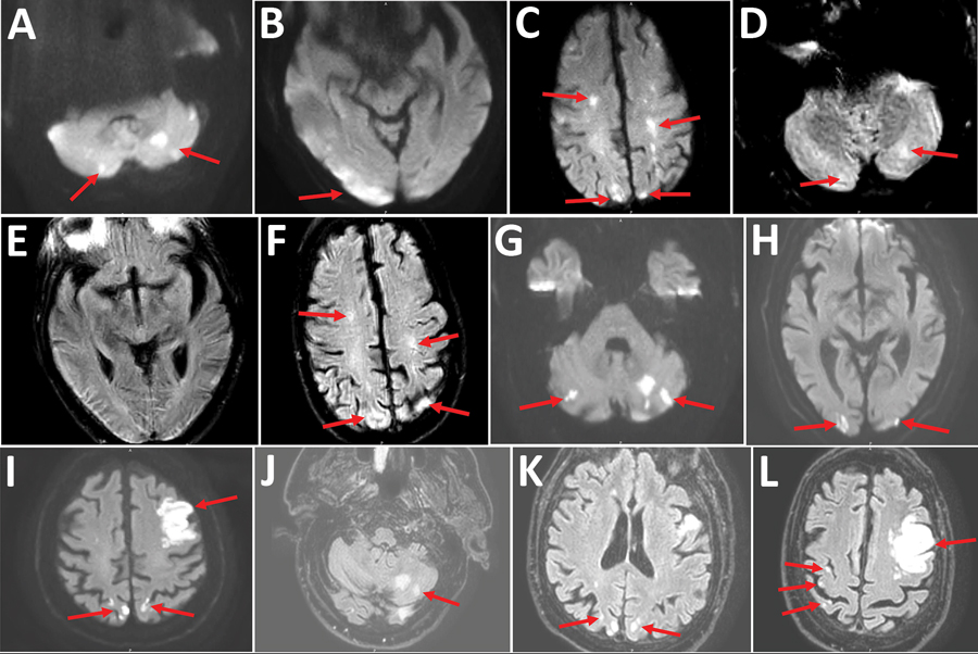
Figure. Cerebral magnetic resonance image (MRI) showing acute ischemic stroke in multiple vascular areas of 2 coronavirus disease patients, France. A–F) Patient 1. Diffusion weighted imaging (DWI) showed hyperintensive lesions of bilateral cerebellar hemispheres (arrows, A), right occipital cortex (arrows, B), bilateral centrum semiovale and bilateral parietal cortex (arrows, C). A part of the lesions are already hyperintensive in FLAIR (fluid-attenuated inversion recovery) sequences (arrows, D, F). Normal FLAIR sequence of the right occipital cortex; early stroke MRI (E). MRI quality is reduced because of dental artifact. G–L) Patient 2. Cerebral MRI showed multiple small ischemic infarctions with hyperintensive lesions (arrows) in bilateral cerebellar hemispheres (DWI [G], FLAIR [J; only left hemisphere]), bilateral occipital cortex (DWI [H], FLAIR [K]), main infarction in the left frontal lobe and small biparietal infarctions (DWI [I], FLAIR [L]).