Volume 27, Number 1—January 2021
Synopsis
Aspergillosis Complicating Severe Coronavirus Disease
Kieren A. Marr , Andrew Platt, Jeffrey A. Tornheim, Sean X. Zhang, Kausik Datta, Celia Cardozo, and Carolina Garcia-Vidal
Figure 1
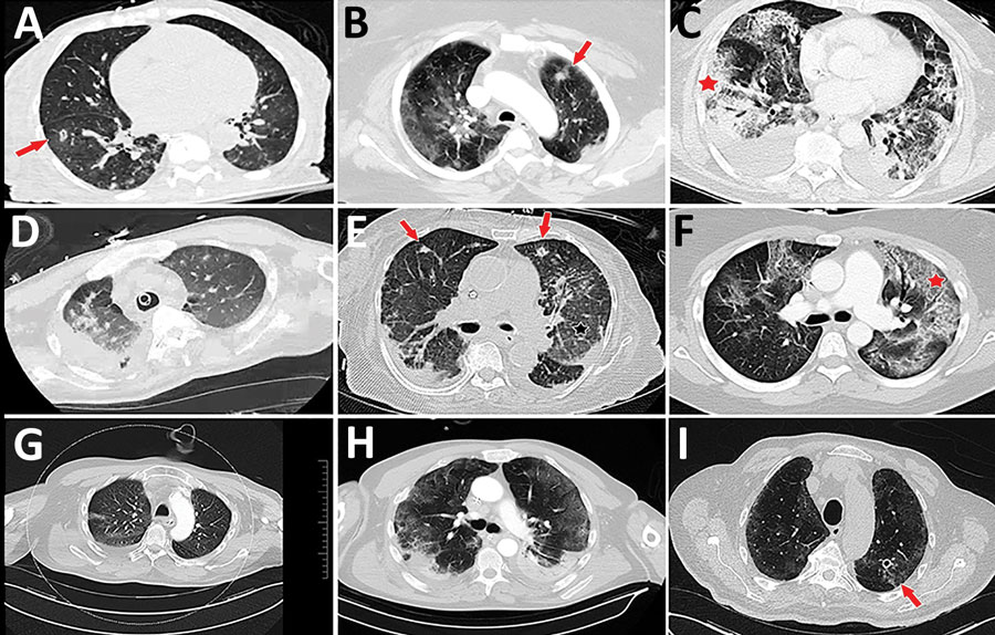
Figure 1. Representative computed tomography (CT) scans for 9 patients with aspergillosis complicating severe viral pneumonia in patients with coronavirus disease. Scans were obtained at or around diagnosis of coronavirus disease–associated pulmonary aspergillosis in this series of patients, described in the Table (https://wwwnc.cdc.gov/EID/article/27/1/20-2896-T1.htm). Corresponding case-patients are indicated with lettered superscripts in the radiology column of Table 1. Examples of nodules and cavitating nodules are indicated by red arrows, and prominent airway thickening and bronchiectasis in ground glass opacities are indicated by red stars.
Page created: September 25, 2020
Page updated: December 21, 2020
Page reviewed: December 21, 2020
The conclusions, findings, and opinions expressed by authors contributing to this journal do not necessarily reflect the official position of the U.S. Department of Health and Human Services, the Public Health Service, the Centers for Disease Control and Prevention, or the authors' affiliated institutions. Use of trade names is for identification only and does not imply endorsement by any of the groups named above.