Skip directly to site content Skip directly to page options Skip directly to A-Z link Skip directly to A-Z link Skip directly to A-Z link
Volume 27, Number 10—October 2021
Dispatch

Widespread Disease in Hedgehogs (Erinaceus europaeus) Caused by Toxigenic Corynebacterium ulcerans

An MartelComments to Author , Filip Boyen, Jörg Rau, Tobias Eisenberg, Andreas Sing, Anja Berger, Koen Chiers, Sarah Van Praet, Serge Verbanck, Muriel Vervaeke, and Frank Pasmans
Author affiliations: Ghent University, Merelbeke, Belgium (A. Martel, F. Boyen, K. Chiers, S. Van Praet, S. Verbanck, F. Pasmans); Chemical and Veterinary Analysis Agency Stuttgart, Fellbach, Germany (J. Rau); Hessian State Laboratory, Giessen, Germany (T. Eisenberg); Bavarian Health and Food Safety Authority, Oberschleißheim, Germany (A. Sing, A. Berger); Agency for Nature and Forests, Brussels, Belgium (M. Vervaeke)

Main Article

Figure 1

Toxigenic Corynebacterium ulcerans as cause of widespread disease in hedgehogs (Erinaceus europaeus), Flanders, Belgium. A) Locations of 81 hedgehogs with lesions on the head or limbs from 4 regions in Flanders, Belgium, who were tested for C. ulcerans. Green, blue, black, and yellow indicate proportion of positive animals; gray indicates proportion of negative animals. Regions: A, Geraardsbergen; B, Merelbeke; C, Herenthout; D, Oudsbergen. Scale bar = 50 km. B) Representative clinical state with necrotizing facial dermatitis in 1 male hedgehog from Merelbeke. C) Ulcerative dermatitis with suppurative exudation and inflammation extending into the subcutis and underlying skeletal muscles. Hematoxylin and eosin stained; scale bar = 100 μm. D) Microcolonies of gram-positive bacilli in suppurative exudate. Gram stain; scale bar = 10 μm.

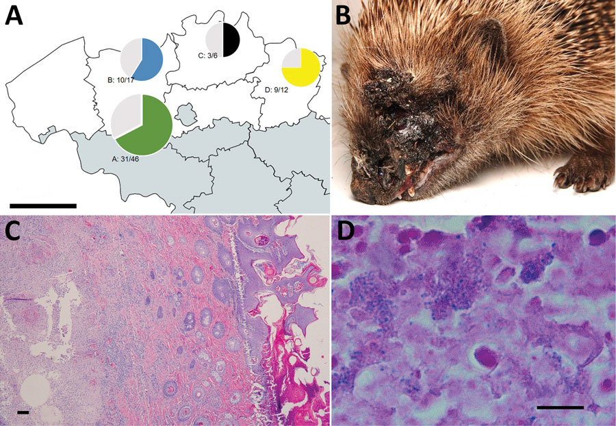
Figure 1. Toxigenic Corynebacterium ulcerans as cause of widespread disease in hedgehogs (Erinaceus europaeus), Flanders, Belgium. A) Locations of 81 hedgehogs with lesions on the head or limbs from 4 regions in Flanders, Belgium, who were tested for C. ulcerans. Green, blue, black, and yellow indicate proportion of positive animals; gray indicates proportion of negative animals. Regions: A, Geraardsbergen; B, Merelbeke; C, Herenthout; D, Oudsbergen. Scale bar = 50 km. B) Representative clinical state with necrotizing facial dermatitis in 1 male hedgehog from Merelbeke. C) Ulcerative dermatitis with suppurative exudation and inflammation extending into the subcutis and underlying skeletal muscles. Hematoxylin and eosin stained; scale bar = 100 μm. D) Microcolonies of gram-positive bacilli in suppurative exudate. Gram stain; scale bar = 10 μm.

Main Article

Page created: June 28, 2021
Page updated: September 19, 2021
Page reviewed: September 19, 2021
The conclusions, findings, and opinions expressed by authors contributing to this journal do not necessarily reflect the official position of the U.S. Department of Health and Human Services, the Public Health Service, the Centers for Disease Control and Prevention, or the authors' affiliated institutions. Use of trade names is for identification only and does not imply endorsement by any of the groups named above.
file_external