Volume 27, Number 10—October 2021
Research
Fatal Cowpox Virus Infection in Human Fetus, France, 2017
Audrey Ferrier1, Gaelle Frenois-Veyrat1, Evelyne Schvoerer, Sandrine Henard, Fanny Jarjaval, Isabelle Drouet, Hawa Timera, Laetitia Boutin, Estelle Mosca, Christophe Peyrefitte, and Olivier Ferraris
Figure 3
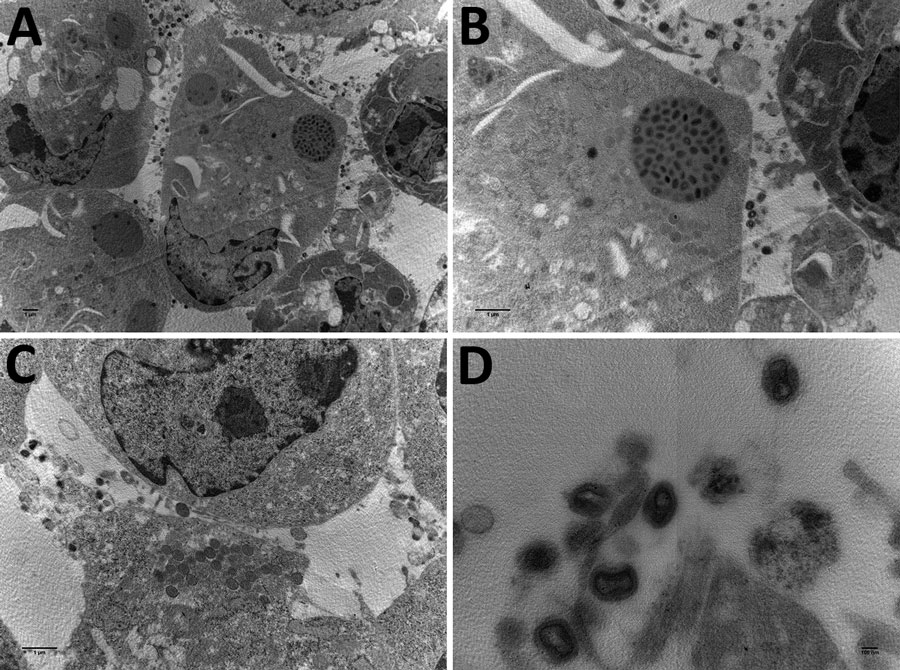
Figure 3. Electron microscopy images of cowpox virus CPXV-54-1716-France (CPXV-like 2), obtained from a pregnant woman in France, 2017. A) Ultrathin sections of BHK-21 cell at 42 hours after infection. Arrow indicates a typical inclusion in the cell cytoplasm. Original magnification ×4,600. B) Higher magnification of BHK-21 cell in panel A. Original magnification ×46,000. C) Ultrathin section of a BHK-21 cell with typical viral factories near the nucleus. Arrows indicate incomplete viruses. Original magnification ×10,500. D) Extracellular-enveloped viruses (arrow). Original magnification ×10,500.
1These authors are co–first authors.
Page created: July 30, 2021
Page updated: September 19, 2021
Page reviewed: September 19, 2021
The conclusions, findings, and opinions expressed by authors contributing to this journal do not necessarily reflect the official position of the U.S. Department of Health and Human Services, the Public Health Service, the Centers for Disease Control and Prevention, or the authors' affiliated institutions. Use of trade names is for identification only and does not imply endorsement by any of the groups named above.