Volume 27, Number 2—February 2021
Research Letter
Intrauterine Transmission of SARS-CoV-2
Figure

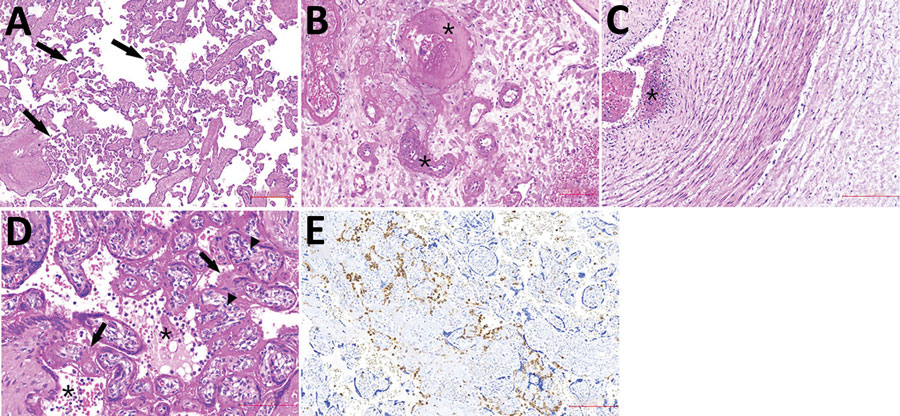
Figure. Histologic sections from the placenta of stillborn fetus of a woman with severe acute respiratory syndrome coronavirus 2 infection, Brazil, 2020. Tissue stained with hematoxylin and eosin. A) Placenta shows accelerated villous maturation with increase in syncytial knots. Black arrows indicate small or short hyper mature villi. B) Membranes and basal decidua show decidual arteriopathy, including fibrinoid necrosis with foam cells, mural hypertrophy, absence of spiral artery remodeling, and arterial thrombosis associated with decidual infarct. Asterisks (*) indicate fibrinoid necrosis. C) The umbilical cord shows subendothelial edema and nonocclusive arterial thrombosis, which was also focally observed in a chorionic plate and stem vessels. Asterisks (*) indicate arterial thrombosis. D–E) Photomicrographs show diffuse perivillous fibrin deposition associated with multifocal mononuclear inflammatory infiltrate in the intervillous space and occasional intervillous thrombi. Black arrows indicate fibrin deposition; asterisks (*) indicate mononuclear infiltrate; arrowheads indicate increase in number of Hofbauer cells. E) Immunohistochemical assay using CD68 antibodies highlights histiocyte infiltrate in paraffin-embedded samples (KP1 Clone; Biocare Medical LLC, https://biocare.net).
1These first authors contributed equally to this article.