Volume 27, Number 2—February 2021
Research Letter
Protective Immunity and Persistent Lung Sequelae in Domestic Cats after SARS-CoV-2 Infection
Figure 1
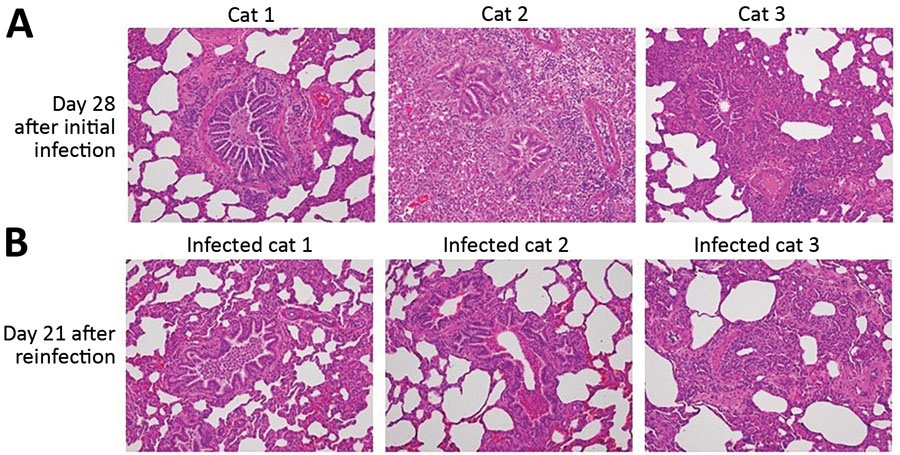
Figure 1. Comparison of histopathology between cats on day 28 after initial infection with severe acute respiratory syndrome coronavirus 2 and on day 21 after reinfection. Bronchioles and alveoli of cats (cats 1–3 in Appendix Figure 6) on day 28 after initial infection (A) and those of cats (infected cats 1–3 in Appendix Figure 6, upper half) on day 21 after reinfection (49 days after the initial infection) (B); original magnification × 20. Cats from both groups showed histiocytic bronchiolitis with occlusive plugs, peribronchiolar fibrosis, and thickening of alveolar septa. Mild acute hemorrhage was detected in affected and less affected regions of the lung on day 21 after reinfection, with a trend toward an increase compared with day 28 (severity score 1.8 + SEM 0.8 on day 21 vs. 0.3 + SEM 0.2 on day 28; p = 0.187 by unpaired t-test).