Skip directly to site content Skip directly to page options Skip directly to A-Z link Skip directly to A-Z link Skip directly to A-Z link
Volume 27, Number 4—April 2021
Dispatch

Genomic Analysis of Novel Poxvirus Brazilian Porcupinepox Virus, Brazil, 2019

Aline S. HoraComments to Author , Sueli A. Taniwaki, Nathana B. Martins, Nataly N.R. Pinto, André E. Schlemper, André L.Q. Santos, Matias P.J. Szabó, and Paulo E. Brandão
Author affiliations: Federal University of Uberlândia, Minas Gerais, Brazil (A.S. Hora, N.B. Martins, N.N.R. Pinto, A.E. Schlemper, A.L.Q. Santos, M.P.J. Szabó); University of São Paulo, São Paulo, Brazil (S.A. Taniwaki, P.E. Brandão)

Main Article

Figure 1

Photographs and histopathology of Brazilian porcupine (Coendou prehensilis) with novel poxvirus tentatively named Brazilian porcupinepox virus, Brazil, 2019. A) Severely swollen and erythematous skin of the eyelids, nasal region, and around oral cavity. B) Severely swollen skin of the forelimbs. C) Histopathologic examination of skin. Marked epidermal hyperplasia and swollen epithelial cells with foci of ballooning degeneration are marked with the square, and parakeratotic hyperkeratosis is indicated by the line. Dermal hemorrhage at the dermal–epidermal junction is indicated with the oval. Hematoxylin and eosin stain. Scale bar indicates 200 μm. D) Histopathologic examination of skin. Cytoplasm of several epithelial cells of epidermis with round eosinophilic inclusions is indicated by arrows. Hematoxylin and eosin stain. Scale bar indicates 200 μm.

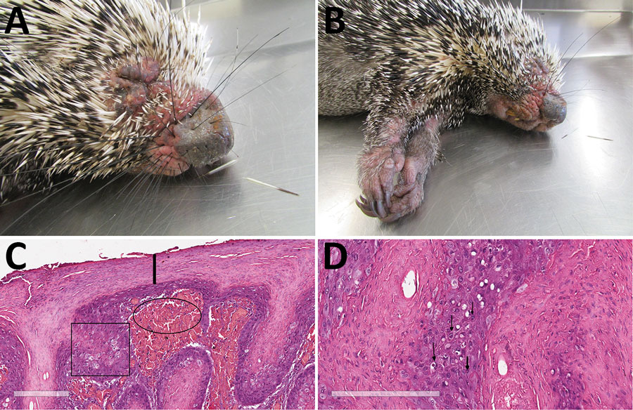
Figure 1. Photographs and histopathology of Brazilian porcupine (Coendou prehensilis) with novel poxvirus tentatively named Brazilian porcupinepox virus, Brazil, 2019. A) Severely swollen and erythematous skin of the eyelids, nasal region, and around oral cavity. B) Severely swollen skin of the forelimbs. C) Histopathologic examination of skin. Marked epidermal hyperplasia and swollen epithelial cells with foci of ballooning degeneration are marked with the square, and parakeratotic hyperkeratosis is indicated by the line. Dermal hemorrhage at the dermal–epidermal junction is indicated with the oval. Hematoxylin and eosin stain. Scale bar indicates 200 μm. D) Histopathologic examination of skin. Cytoplasm of several epithelial cells of epidermis with round eosinophilic inclusions is indicated by arrows. Hematoxylin and eosin stain. Scale bar indicates 200 μm.

Main Article

Page created: February 02, 2021
Page updated: March 18, 2021
Page reviewed: March 18, 2021
The conclusions, findings, and opinions expressed by authors contributing to this journal do not necessarily reflect the official position of the U.S. Department of Health and Human Services, the Public Health Service, the Centers for Disease Control and Prevention, or the authors' affiliated institutions. Use of trade names is for identification only and does not imply endorsement by any of the groups named above.
file_external