Volume 27, Number 4—April 2021
Synopsis
Difficulties in Differentiating Coronaviruses from Subcellular Structures in Human Tissues by Electron Microscopy
Hannah A. Bullock , Cynthia S. Goldsmith, Sherif R. Zaki, Roosecelis B. Martines, and Sara E. Miller
Figure 3
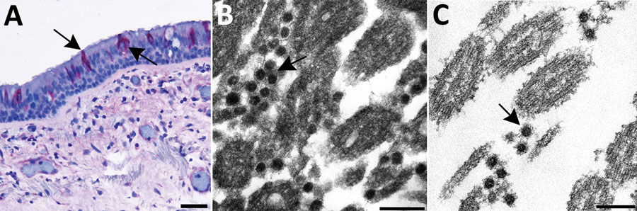
Figure 3. Use of immunohistochemistry and electron microscopy to detect severe acute respiratory syndrome coronavirus 2 (SARS-CoV-2) in formalin-fixed paraffin embedded (FFPE) autopsy tissues. A) Immunostaining (arrows) of SARS-CoV-2 in the epithelial cells of the trachea. Scale bar indicates 20 µm. B) Ultrastructural features of extracellular SARS-CoV-2 particles (arrow) in association with ciliated cells of the trachea from paraffin section in panel A, prepared using an FFPE on-slide method. Scale bar indicates 200 nm. C) Thin section of a biopsy punch from the original FFPE block in panel A showing viral particles (arrow) ≈75 nm. Scale bar indicates 200 nm.
Page created: February 18, 2021
Page updated: March 18, 2021
Page reviewed: March 18, 2021
The conclusions, findings, and opinions expressed by authors contributing to this journal do not necessarily reflect the official position of the U.S. Department of Health and Human Services, the Public Health Service, the Centers for Disease Control and Prevention, or the authors' affiliated institutions. Use of trade names is for identification only and does not imply endorsement by any of the groups named above.