Volume 27, Number 5—May 2021
Research Letter
Eosinophilic Meningitis and Intraocular Infection Caused by Dirofilaria sp. Genotype Hongkong
Cite This Article
Citation for Media
Abstract
Eosinophilic meningitis caused by human diroflarial infection is rare. We report a case of eosinophilic meningitis and concomitant intraocular dirofilarial infection in India. Sequencing of the mitochondrial genome identified the worm as Dirofilaria sp. genotype Hongkong, a close relative of D. repens nematodes.
Dirofilariasis is a group of mosquitoborne parasitoses. The most prevalent Dirofilaria species causing infection are D. imitis and D. repens nematodes (1). Dogs are the definitive hosts in the life cycle, in which microfilaremia is observed. Humans are aberrant hosts, and the worms usually remain infertile (1,2). Human dirofilariasis is reported mostly as 1 worm in the subconjunctival or subcutaneous spaces. Surgical extraction of the worm constitutes definitive therapy. These worms are rarely observed inside the eye (1,2). Identification of the worm by using morphologic features is difficult because a large number of Dirofilaria species have similar features.
Diagnosis of eosinophilic meningitis is based mainly on clinical features and microscopic identification of eosinophils in the central nervous system. Helminthic infections, such as angiostrongylosis, baylisascariasis, and gnathostomiasis, are most commonly implicated in eosinophilic meningitis (3). We report a rare case of eosinophilic meningitis and concomitant intraocular dirofilarial infection. Sequencing of the mitochondrial genome of the extracted worm identified it as Dirofilaria sp. genotype Hongkong, a close relative of D. repens (4).
A 17-year-old woman came to our institute in Kochi, India, because of acute onset of severe headache, irritability, visual blurring, and diplopia, after 3 weeks of intermittent fever. She had meningeal signs, bilateral lateral rectus palsy, and papilledema. Peripheral eosinophilia (14.2%) was observed. Magnetic resonance imaging of the brain (Appendix Figure 1) showed diffuse leptomeningeal enhancement. Cerebrospinal fluid showed lymphocytic pleocytosis (1,040 cells/μL), major eosinophilia (37%), and protein and glucose levels within reference ranges.
Figure
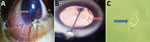
Figure. Eosinophilic meningitis and intraocular infection caused by Dirofilariasp. genotype Hongkong in a patient in Kochi, Inida. A) Organism (arrow) in the left eye of patient during routine clinical...
Video
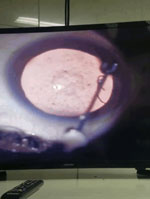
Video. Live, mobile worm in anterior chamber of left eye of the patient. Video was taken while lignocaine was being injected.
A live worm was detected in the anterior chamber of her left eye (Figure, panel A), confirmed by slit lamp examination (Figure, panel B; Video). The lens showed cataractous changes. Indirect ophthalmoscopy showed inflammatory changes in retinal pigment epithelium, suggestive of a migratory tract. Serologic analysis for helminthic antibodies was not conducted because serologic testing was were not available. A white, thread-like worm (length ≈15 mm) was extracted after the worm was paralyzed by injection of lignocaine into the anterior chamber of the eye (Figure, panel C).
Because a PCR was available, histopathologic analysis was not conducted. Morphologic features or sex could not be determined. The worm specimen was subjected to multiplex PCR for D. repens and D. imitis using an equimolar combination of general and species-specific primers: Diro_12S_F (5′-GTTCCAGAATAATCGGCTA-3′), Diro_12S_R (5′-ATTGACGGATGGTTTGTACC-3′), D. immitis_F (5′-TTTTTACTTTTTTGGTAATG-3′), and D. repens_R (5′-AAAAGCAACACAAATAAAA-3′). The cytochrome c oxidase subunit 1 (COX1) region was amplified by using primers Fil_COX1F (5′-GCTTTRTCTTTTTGGKTTACTTTT-3′) and Fil_COX1R (5′-TAGTRTCATAAAAAGAAGTATTAAA-3′) (5).
Although the specimen was identified as a D. repens worm, Sanger sequencing of the COX1 and 12S rDNA PCR products was performed by using the BigDye Terminator v3.1 Cycle Sequencing Kit (Applied Biosystems, https://www.thermofisher.com) and the Genetic Analyzer 3130XL (Applied Biosystems). Sequences of 12S rRNA and COXI genes obtained were deposited in GenBank (accession nos. MT984272 and MT984209).
Phylogenetic analysis of the 12S rRNA (MT984272) and COX1 (MT984209) sequences obtained from the isolate was performed by using the maximum-likelihood method with 1,000 bootstrap replications and MEGA X version 7 (https://www.megasoftware.net). Both the 12S rRNA and the COX1 sequences obtained from the human isolate were in the same cluster with Dirofilaria sp. genotype Hongkong and were separated from other Dirofilaria species (5,6) (Appendix Figure 2). Peripheral blood smears were negative for microfilaria. Symptoms of the patient resolved slowly after worm extraction and initiation of treatment with steroids.
Migrating worms in humans might cause a variety of clinical problems, which could be caused by mechanical effects or immune responses. Intraocular parasites might induce severe damage to various structures in the eye. Literature on eosinophilic meningitis and concomitant ocular parasites is limited. Clinical manifestations of eosinophilic meningitis are usually attributed to the severe inflammatory response incited by migrating worms, even though they are rarely demonstrated in vivo. Eosinophilic meningitis caused by Angiostrongylus cantonensis worms has been frequently reported in the Asia–Pacific region (7). Dirofilaria infection rarely results in eosinophilic meningitis (1,2).
Poppert et al. reported a case of D. repens infection, which was subsequently identified as Dirofilaria sp. genotype Hongkong, which caused subcutaneous infection and concomitant eosinophilic meningoencephalitis in a traveler returning from Kerala, India, and Sri Lanka to Germany (8). Subconjunctival infection with Dirofilaria sp. genotype Hongkong has also been reported in a patient returning to Austria after a 7-week stay in India (9). A recent study from Kerala, India, suggested that most of D. repens infections reported from southern India have the Dirofilaria sp. Hongkong genotype (10).
Demonstration of a live, intraocular worm and its subsequent identification as Dirofilaria sp. genotype Hongkong by using sequencing added a new dimension to this case of eosinophilic meningitis. Infection with the Dirofilaria sp. Hongkong genotype, blood eosinophilia, and eosinophilic meningitis are the 3 strikingly similar features between our case-patient and Poppert et al. (8), suggesting that Dirofilaria sp. genotype Hongkong might induce a more systemic eosinophilic reaction than D. repens.
Sequencing using panfilarial primers might help characterize most filarial species. Such an approach might clarify the etiopathogenesis of eosinophilic meningitis, leading to newer therapeutic and preventive strategies.
Dr. Jyotsna is a resident in pediatric neurology at Amrita Institute of Medical Sciences and Research Centre, Kochi, Kerala, India. Her primary research interest is neurologic manifestations of infectious diseases.
References
- Genchi C, Rinaldi L, Mortarino M, Genchi M, Cringoli G. Climate and Dirofilaria infection in Europe. Vet Parasitol. 2009;163:286–92. DOIPubMedGoogle Scholar
- Simón F, Siles-Lucas M, Morchón R, González-Miguel J, Mellado I, Carretón E, et al. Human and animal dirofilariasis: the emergence of a zoonotic mosaic. Clin Microbiol Rev. 2012;25:507–44. DOIPubMedGoogle Scholar
- Graeff-Teixeira C, da Silva AC, Yoshimura K. Update on eosinophilic meningoencephalitis and its clinical relevance. Clin Microbiol Rev. 2009;22:322–48. DOIPubMedGoogle Scholar
- To KK, Wong SS, Poon RW, Trendell-Smith NJ, Ngan AH, Lam JW, et al. A novel Dirofilaria species causing human and canine infections in Hong Kong. J Clin Microbiol. 2012;50:3534–41. DOIPubMedGoogle Scholar
- Gioia G, Lecová L, Genchi M, Ferri E, Genchi C, Mortarino M. Highly sensitive multiplex PCR for simultaneous detection and discrimination of Dirofilaria immitis and Dirofilaria repens in canine peripheral blood. Vet Parasitol. 2010;172:160–3. DOIPubMedGoogle Scholar
- To KK, Wong SS, Poon RW, Trendell-Smith NJ, Ngan AH, Lam JW, et al. A novel Dirofilaria species causing human and canine infections in Hong Kong. J Clin Microbiol. 2012;50:3534–41. DOIPubMedGoogle Scholar
- Wang QP, Wu ZD, Wei J, Owen RL, Lun ZR. Human Angiostrongylus cantonensis: an update. Eur J Clin Microbiol Infect Dis. 2012;31:389–95. DOIPubMedGoogle Scholar
- Poppert S, Hodapp M, Krueger A, Hegasy G, Niesen WD, Kern WV, et al. Dirofilaria repens infection and concomitant meningoencephalitis. Emerg Infect Dis. 2009;15:1844–6. DOIPubMedGoogle Scholar
- Winkler S, Pollreisz A, Georgopoulos M, Bagò-Horvath Z, Auer H, To KK, et al. Candidatus Dirofilaria hongkongensis as causative agent of human ocular filariosis after travel to India. Emerg Infect Dis. 2017;23:1428–31. DOIPubMedGoogle Scholar
- Pradeep RK, Nimisha M, Pakideery V, Johns J, Chandy G, Nair S, et al. Whether Dirofilaria repens parasites from South India belong to zoonotic Candidatus Dirofilaria hongkongensis (Dirofilaria sp. hongkongensis)? Infect Genet Evol. 2019;67:121–5. DOIPubMedGoogle Scholar
Figures
Cite This ArticleOriginal Publication Date: April 13, 2021
Table of Contents – Volume 27, Number 5—May 2021
| EID Search Options |
|---|
|
|
|
|
|
|
Please use the form below to submit correspondence to the authors or contact them at the following address:
Kollencheri P. Vinayan, Department of Pediatric Neurology, Amrita Institute of Medical Sciences and Research Centre, Ponnekkara, Edapally, Kochi 682041 India
Top