Volume 27, Number 5—May 2021
Research Letter
Intersecting Paths of Emerging and Reemerging Infectious Diseases
Figure

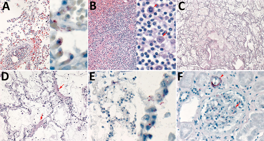
Figure. Histopathologic and immunohistochemical characteristics of fatal hantavirus pulmonary syndrome in 2 patients, Arizona, USA, 2020. A) Patient 1 lung tissue, showing intravascular leukocytosis with left shift (left, original magnification ×50) and hantavirus antigen immunostaining (red) in pulmonary microvasculature (right, original magnification ×158). B) Patient 1 spleen tissue, showing immunoblast proliferation in the red pulp and periarteriolar sheaths (left, original magnification ×50) and immunoblasts with high nuclear to cytoplasmic ratio, vesicular and prominent nucleoli (arrows) and mitosis (arrowhead) (right, original magnification ×158). C) Patient 2 lung tissue, showing severe intraalveolar edema (original magnification ×12.5). D) Patient 2 lung tissue, showing interstitial pneumonitis with hyaline membranes (arrows) (original magnification ×50). E) Patient 2 lung tissue, showing hantavirus antigen immunostaining (red) in pulmonary microvasculature (left, original magnification ×50; right, original magnification ×158). F) Patient 2 kidney tissue, showing hantavirus antigen immunostaining (red) in glomerular capillaries (arrowhead) and interstitial vessel (arrow) (original magnification ×100).