Volume 27, Number 6—June 2021
Dispatch
Recurrent Swelling and Microfilaremia Caused by Dirofilaria repens Infection after Travel to India
Figure 1
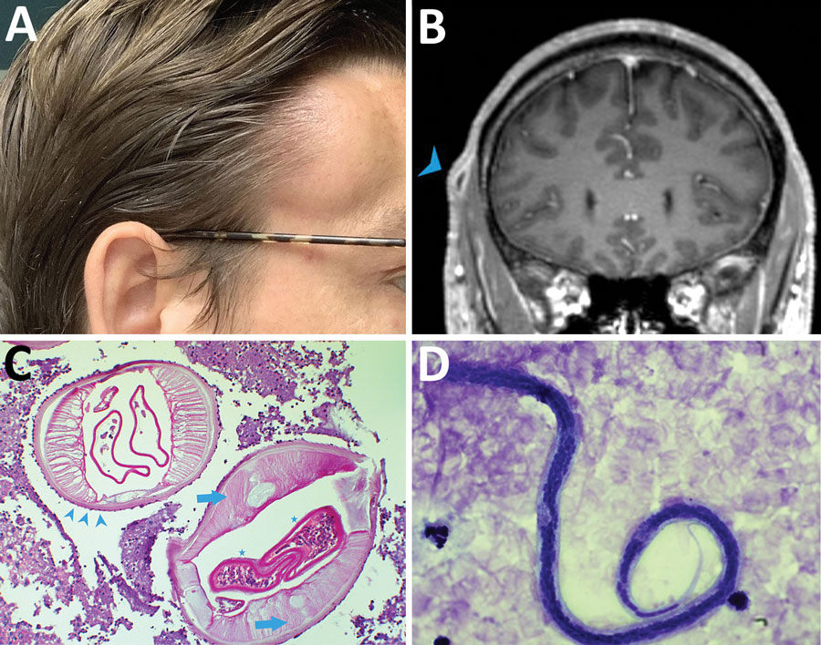
Figure 1. Dirofilaria repens infection in man in Germany after travel to India. A) Painless temporal subcutaneous swelling (image taken by the patient at the time of maximal protrusion). B) Contrast-enhanced magnetic resonance image (fat-saturated T1-weighted sequences) demonstrating a subcutaneous 10-mm lesion with central hypointensity and contrast uptake of the surrounding capsule (arrowhead). C) Cross-section through adult D. repens worm in subcutaneous tissue, demonstrating the cuticle with external ridges (arrow heads) and internal structures such as smooth muscle fibers (arrows) and gravid uteri (stars). Original magnification ×100; periodic acid-Schiff stain. D) D. repens microfilaria of the Asian genotype. Typical features include lack of a sheath, 2–3 separate nuclei in the head space, and absence of nuclei in the tip of the tail. Original magnification ×1,000 with oil; Giemsa stain.
1These senior authors contributed equally to this article.