Volume 27, Number 7—July 2021
Research
Novel Morbillivirus as Putative Cause of Fetal Death and Encephalitis among Swine
Figure 2
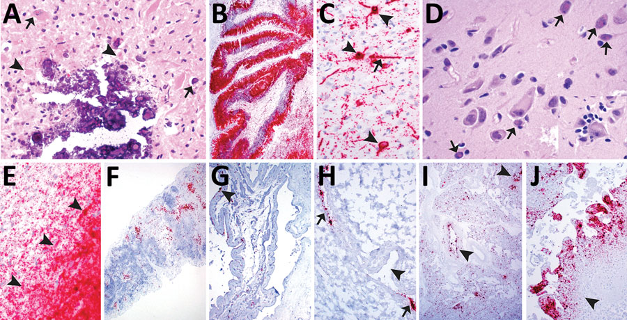
Figure 2. Histologic lesions and porcine morbillivirus (PoMV) RNA in situ hybridization (ISH, red) of tissue of infected swine. A) Histologic section of cerebrum from fetus A stained by hematoxylin and eosin. Arrowheads indicate neuronal necrosis; arrows indicate mineralization and viral inclusion bodies in a neuron and glial cell. B) Cerebellum of fetus A with extensive detection of PoMV by ISH. C) Cerebrum of fetus A; arrowheads indicate ISH labeling within the cytoplasmic and nuclear compartment of neurons; arrow indicates ISH labelling in an axon. D) Cerebrum of fetus B; arrows indicate multiple viral inclusion bodies in neurons; inset displays satellitosis. E) Cerebrum of fetus B showing extensive PoMV detection by ISH. Arrowheads indicate the border of white and gray matter. F) Detection of PoMV by ISH in the spleen of fetus C. G) Detection of PoMV by ISH in a placenta from litter D; arrowhead indicates allantoic epithelium. H) Detection of PoMV by ISH in a renal vessel of a fetus from litter D; arrows indicate the endothelium and arrowhead indicates the vessel lumen. I) Detection of PoMV by ISH in conducting airways (arrowheads) and alveolar septa in the lung of fetus from litter E. J) Detection of PoMV by ISH in the allantoic connective tissue of the placenta and leukocytes from litter F; arrowhead indicates infiltration of leukocytes.