Volume 27, Number 8—August 2021
Synopsis
Mycobacterium microti Infections in Free-Ranging Red Deer (Cervus elaphus)
Giovanni Ghielmetti , Anne M. Kupca, Matthias Hanczaruk, Ute Friedel, Hubert Weinberger, Sandra Revilla-Fernández, Erwin Hofer, Julia M. Riehm, Roger Stephan, and Walter Glawischnig
Figure 2
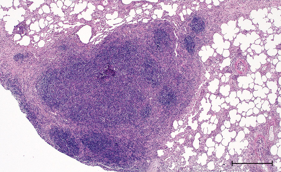
Figure 2. Histopathologic features in red deer in case 2 in study of tuberculosis caused by Mycobacterium microti in red deer, Austria and Germany. Lung tissue highly infiltrated by round cells, predominantly lymphocytes and some macrophages, single multinucleated Langhans-type giant cells, hematoxylin and eosin stain. Scale bar = 500 μm.
Page created: May 12, 2021
Page updated: July 18, 2021
Page reviewed: July 18, 2021
The conclusions, findings, and opinions expressed by authors contributing to this journal do not necessarily reflect the official position of the U.S. Department of Health and Human Services, the Public Health Service, the Centers for Disease Control and Prevention, or the authors' affiliated institutions. Use of trade names is for identification only and does not imply endorsement by any of the groups named above.