Volume 27, Number 9—September 2021
Research
Multicenter Epidemiologic Study of Coronavirus Disease–Associated Mucormycosis, India
Atul Patel1, Ritesh Agarwal12, Shivaprakash M. Rudramurthy, Manoj Shevkani, Immaculata Xess, Ratna Sharma, Jayanthi Savio, Nandini Sethuraman, Surabhi Madan, Prakash Shastri, Deepak Thangaraju, Rungmei Marak, Karuna Tadepalli, Pratik Savaj, Ayesha Sunavala, Neha Gupta, Tanu Singhal, Valliappan Muthu, Arunaloke Chakrabarti2 , and MucoCovi Network3
Figure 4
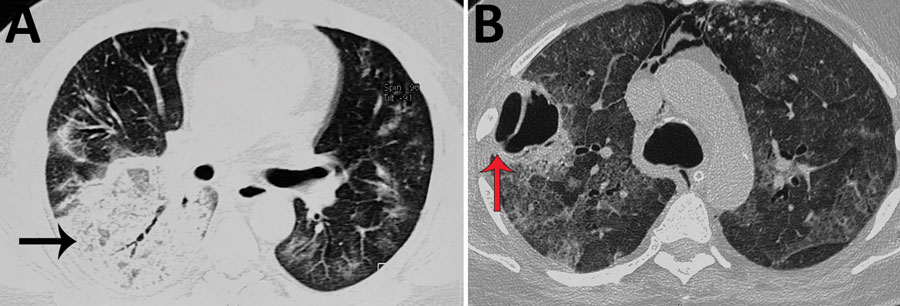
Figure 4. Noncontrast computed tomography scan of the thorax of a patient with coronavirus disease–associated mucormycosis, India, 2020. A) Pulmonary mucormycosis demonstrated as a large area of consolidations with patchy air trapping (black arrow), patchy ground-glass opacities, and septal thickening; B) large thick-walled cavity (red arrow) with surrounding ground-glass opacities.
1These first authors contributed equally to this article.
2These senior authors contributed equally to this article.
3Members are listed at the end of this article.
Page created: June 02, 2021
Page updated: August 17, 2021
Page reviewed: August 17, 2021
The conclusions, findings, and opinions expressed by authors contributing to this journal do not necessarily reflect the official position of the U.S. Department of Health and Human Services, the Public Health Service, the Centers for Disease Control and Prevention, or the authors' affiliated institutions. Use of trade names is for identification only and does not imply endorsement by any of the groups named above.