Dirofilaria repens Testicular Infection in Child, Italy
Sara Ugolini

, Mario Lima, Michela Maffi, Francesco Pierangeli, Marzia Vastano, Tommaso Gargano, Stefania Varani, Andrea Gustinelli, Monica Caffara, and Maria L. Fioravanti
Author affiliations: Wythenshawe Hospital, Manchester, UK (S. Ugolini); University of Manchester NHS Foundation Trust, Manchester (S. Ugolini); IRCCS Azienda Ospedaliero–Universitaria di Bologna, Bologna, Italy (M. Lima, M. Maffi, M. Vastano, T. Gargano, S. Varani); Ospedali Riuniti di Ancona, Ancona, Italy (F. Pierangeli); Alma Mater Studiorum–University of Bologna, Bologna (M. Lima, T. Gargano, S. Varani, A. Gustinelli, M. Caffara, M.L. Fioravanti)
Main Article
Figure
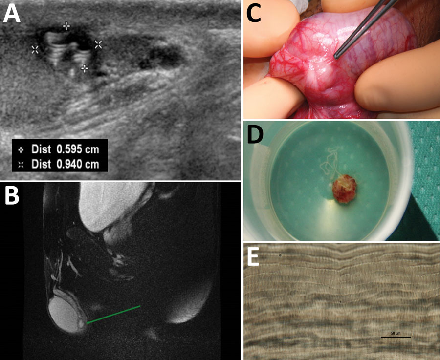
Figure. Diagnostic evaluation of Dirofilaria repens testicular infection in a child from Italy, a boy 13 years of age who had a 5-month history of swelling in the left testicle. A) Ultrasound scan showed a 0.5 × 0.9 cm hypoechoic cyst with moving artifacts and thread-like hyperechoic structures. B) Magnetic resonance imaging showed the cyst was located on the testis without signs of infiltration and contained fluid mixed with tubular structures and moving artifacts. C) Exploration of the scrotum before cyst excision showed a well-circumscribed, encapsulated tense nodule on the left side. D) The cyst was excised and a coiled roundworm was found in the opened capsule. E) We identified the nematode as a female D. repens nematode by microscopically observing typical longitudinal ridges on the body surface. Scale bar indicates 50 μm. Dist, distance.
Main Article
Page created: September 26, 2022
Page updated: November 21, 2022
Page reviewed: November 21, 2022
The conclusions, findings, and opinions expressed by authors contributing to this journal do not necessarily reflect the official position of the U.S. Department of Health and Human Services, the Public Health Service, the Centers for Disease Control and Prevention, or the authors' affiliated institutions. Use of trade names is for identification only and does not imply endorsement by any of the groups named above.