Volume 28, Number 2—February 2022
Research Letter
Fatal Case of Mediterranean Spotted Fever Associated with Septic Shock, Iran
Cite This Article
Citation for Media
Abstract
A fatal case of Mediterranean spotted fever associated with septic shock was reported in a 61-year-old man living in a village in southeastern Iran. The patient had a history of tick bite a few days before symptom onset. Phylogenetic analysis confirmed infection by Rickettsia conorii subspecies israelensis.
Mediterranean spotted fever (MSF) is a zoonotic disease caused by Rickettsia conorii. The main vector of this bacterium is the Rhipicephalus sanguineus tick (1); the main hosts of these ticks are domestic dogs, and humans are incidental hosts (2). MSF is endemic to the Mediterranean, Europe, Africa, western Asia, and India. The case-fatality rate is 3%–7% in hospitalized patients (3,4).
In 2017, human cases of MSF were reported in Kerman province in southeastern Iran (5). No data are available on the epidemiology of MSF in Iran; we report a fatal case of MSF associated with septic shock.
The patient was a 61-year-old man with a 10-year history of hypertension and rheumatoid arthritis who lived in a village in proximity to Bam County, Kerman province, Iran. He was a farmer, had no history of domestic animal-keeping, and reported contact with livestock and a tick bite a few days before symptom onset. The initial clinical signs of the disease appeared on September 6, 2019, and the patient was admitted to a hospital in Bam on September 9; symptoms were fever, nausea, vomiting, myalgia, urinary retention, and flank pain. The patient had scleral icterus, and a black skin eschar at the tick bite site and skin rash were visible on his left leg.
When the patient’s condition deteriorated, he was transferred to a hospital in Kerman on September 15. At admission, symptoms were septic shock, tachycardia, tachypnea, fever, and hypotension (85/50 mm Hg); he immediately began treatment with ceftriaxone, metronidazole, and parenteral hydration. Maculopapular skin rash was visible on the left leg. The patient had thrombocytopenia, and an increase was observed in leukocyte counts, renal factor levels (urea and creatinine), liver enzyme levels (aspartate aminotransferase, alanine transferase, and alkaline phosphatase), partial thromboplastin time of coagulation, and bilirubin levels (Table). Hemoglobin and hematocrit levels decreased, and the patient experienced hematuria and proteinuria; calcium oxalate and amorphous urate crystals were further reported in microscopic examinations. Treatment of prednisolone, heparin, doxycycline, and vancomycin was initiated.
On September 16, the patient was transferred to Afzalipour Hospital in Kerman (Referral Center for Infectious Diseases, Kerman Province). At the time of admission, the patient was conscious, his condition was stable, and his temperature was 37.6°C. No abnormalities were observed in clinical examinations of the heart, chest, and abdomen, but we noted bilateral lower extremity edema and left leg skin lesions (rash and eschar). The results of laboratory tests of blood and urine samples were abnormal (Table). The patient underwent emergency dialysis and continued to take prednisolone, heparin, doxycycline, and vancomycin. On September 17, the patient lost consciousness; he was subsequently intubated and admitted to the intensive care unit. A few hours later, he experienced septic shock and cardiac arrest and died.
Figure
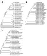
Figure. Phylogenetic analysis of Rickettsia species from a patient with Mediterranean spotted fever associated with septic shock, Iran (in bold), confirming infection with R. conorii subspecies ...
The differential diagnosis for this patient included MSF and Crimean-Congo hemorrhagic fever; on September 17, samples required for these differential diagnoses were prepared. Serum and blood samples were sent to the Pasteur Institute of Iran on September 25 (8 days after the patient’s death). Serologic and molecular test results for Crimean-Congo hemorrhagic fever were negative. Testing for R. conorii IgM by ELISA was borderline, and titer of R. conorii IgM by immunofluorescence assay was 1:48. Serum samples were positive for Rickettsia spp. (16S rRNA gene) by real-time reverse transcription PCR (6). On the basis of the amplification and sequencing of specific genes of Rickettsia spp. (gltA, GenBank accession no. MZ545594.1; 17KD, GenBank accession no. MZ545592.1; ompA, GenBank accession no. MZ545593.1), we confirmed infection by R. conorii subspecies israelensis (Figure).
The patient died as a result of late diagnosis of a rickettsial infection and subsequent septic shock, despite initiation of appropriate treatment. MSF is usually considered to be a mild disease, but severe and fatal cases do occasionally occur (7). One of the causes of death from MSF is multiorgan failure, including acute kidney injury, pneumonitis, and encephalitis. When severe, MSF can manifest as septic shock, and acute kidney injury might occur. Thrombocytopenia and elevated liver enzymes are frequent laboratory abnormalities (4,7).
Phylogenetic trees showed that the infection in this patient was caused by R. conorii subsp. israelensis. R. conorii has 4 subspecies, caspia, israelensis, conorii, and indica, each of which cause diseases that have specific clinical features and occur in different geographic regions. R. conorii subsp. israelensis seems to have the highest death rate of the subspecies (8,9), reported to be ≈30% (10).
MSF appears to be circulating in southern Iran but is a neglected disease that requires more attention from the healthcare system. Because of the nonspecific clinical symptoms of MSF, diagnosing the disease is challenging. Diagnosing and treating MSF early is critical to prevent progression to more severe illness (6). Further studies, particularly on elucidating potential reservoirs and vectors, will result in a better understanding of the epidemiology of this disease in Iran. In the meantime, MSF should be included in the differential diagnosis for patients in Iran who are experiencing fever and rash.
Dr. Esmaeili is an assistant professor and researcher in medical microbiology at Pasteur Institute of Iran. His primary research interest is emerging and reemerging bacterial pathogens, including Coxiella burnetii, Bartonella, Francisella, Rickettsia, and Yersinia pestis.
Acknowledgment
The work presented in this publication was supported by the Pasteur Institute of Iran and the Center for Communicable Diseases Control in the Ministry of Health (grant no. 810).
References
- Rovery C, Brouqui P, Raoult D. Questions on Mediterranean spotted fever a century after its discovery. Emerg Infect Dis. 2008;14:1360–7. DOIPubMedGoogle Scholar
- Nafi O, Tarawnah Y, Tarawnah A. Epidemiological evaluation of Mediterranean spotted fever in children of the Karak province in south Jordan. J Infect Dev Ctries. 2017;11:242–6. DOIPubMedGoogle Scholar
- Parola P, Paddock CD, Raoult D. Tick-borne rickettsioses around the world: emerging diseases challenging old concepts. Clin Microbiol Rev. 2005;18:719–56. DOIPubMedGoogle Scholar
- Baymakova M, Pekova L, Plochev K, Parousheva P. Severe clinical forms of Mediterranean spotted fever: a case series from an endemic area in Bulgaria. Int J Infect Dis. 2016;53:150–1. DOIGoogle Scholar
- Farrokhnia M, Ghalejoogh ZY, Rohani M, Ghasemi A, Esmaeili S, Mostafavi E. Cases of Mediterranean spotted fever in southeast of Iran. Iran J Microbiol. 2020;12:256–60. DOIPubMedGoogle Scholar
- Abdeljelil M, Sakly H, Kooli I, Marrakchi W, Aouam A, Loussaief C, et al. Mediterranean spotted fever as a cause of septic shock. IDCases. 2019;15:
e00528 . DOIPubMedGoogle Scholar - Walker DH, Herrero-Herrero JI, Ruiz-Beltrán R, Bullón-Sopelana A, Ramos-Hidalgo A. The pathology of fatal Mediterranean spotted fever. Am J Clin Pathol. 1987;87:669–72. DOIPubMedGoogle Scholar
- Parola P, Paddock CD, Socolovschi C, Labruna MB, Mediannikov O, Kernif T, et al. Update on tick-borne rickettsioses around the world: a geographic approach. Clin Microbiol Rev. 2013;26:657–702. DOIPubMedGoogle Scholar
- Cohen R, Babushkin F, Shapiro M, Uda M, Atiya-Nasagi Y, Klein D, et al. Two cases of Israeli spotted fever with Purpura fulminans, Sharon District, Israel. Emerg Infect Dis. 2018;24:835–40. DOIPubMedGoogle Scholar
- Sousa R, França A, Dória Nòbrega S, Belo A, Amaro M, Abreu T, et al. Host- and microbe-related risk factors for and pathophysiology of fatal Rickettsia conorii infection in Portuguese patients. J Infect Dis. 2008;198:576–85. DOIPubMedGoogle Scholar
Figure
Table
Cite This ArticleOriginal Publication Date: January 14, 2022
Table of Contents – Volume 28, Number 2—February 2022
| EID Search Options |
|---|
|
|
|
|
|
|
Please use the form below to submit correspondence to the authors or contact them at the following address:
Address for correspondence: Saber Esmaeili, No. 69, Pasteur Ave, Department of Epidemiology and Biostatistics, Pasteur Institute of Iran, Postal Code 1316943551, Tehran, Iran
Top