Skip directly to site content Skip directly to page options Skip directly to A-Z link Skip directly to A-Z link Skip directly to A-Z link
Volume 28, Number 2—February 2022
Research Letter

Probable Transmission of SARS-CoV-2 Omicron Variant in Quarantine Hotel, Hong Kong, China, November 2021

Haogao Gu, Pavithra Krishnan, Daisy Y.M. Ng, Lydia D.J Chang, Gigi Y.Z. Liu, Samuel S.M. Cheng, Mani M.Y. Hui, Mathew C.Y. Fan, Jacob H.L. Wan, Leo H.K. Lau, Benjamin J. Cowling, Malik Peiris, and Leo L.M. PoonComments to Author 
Author affiliation: The University of Hong Kong, Hong Kong, China

Main Article

Figure

Detection of severe acute respiratory syndrome coronavirus 2 Omicron variant in 2 patients (cases A and B) in Hong Kong, China, November 2021. A) Phylogenetic time tree of Omicron nucleotide sequences using an early severe acute respiratory syndrome coronavirus sequence as a reference sequence (Wuhan-Hu-1/2019; GenBank accession no. MN908947.3). B) Comparison of Omicron variant mutations in case A to other variants; red indicates VOC and gray VOI (Appendix). Text colors indicate mutations found in NTD (blue), RBD (orange), FP (purple), and HR1 (green). Lane 1, case A; 2, Alpha (B.1.1.7); 3, Beta (B.1351); 4, Delta (B.1.617.2); 5, Gamma (P1); 6, Epsilon (B.1.427/429); 7, Eta (B.1.525); 8, Iota (B.1.526); 9, Kappa (B.1.617.1); 10, Lambda (C.37); 11, Mu (B.1.1.621); 12, Theta (P.3); 13, Zeta (P.2). E, envelope; FP, fusion peptide; HR1, heptad repeat 1; M, matrix; NSP, nonstructural protein; NTD, N-terminal domain; RBD, receptor-binding domain; S, spike; VOC, variant of concern; VOI, variant of interest.

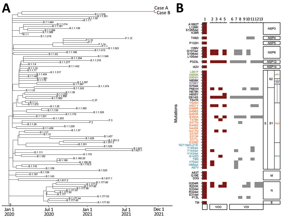
Figure. Detection of severe acute respiratory syndrome coronavirus 2 Omicron variant in 2 patients (cases A and B) in Hong Kong, China, November 2021. A) Phylogenetic time tree of Omicron nucleotide sequences using an early severe acute respiratory syndrome coronavirus sequence as a reference sequence (Wuhan-Hu-1/2019; GenBank accession no. MN908947.3). B) Comparison of Omicron variant mutations in case A to other variants; red indicates VOC and gray VOI (Appendix). Text colors indicate mutations found in NTD (blue), RBD (orange), FP (purple), and HR1 (green). Lane 1, case A; 2, Alpha (B.1.1.7); 3, Beta (B.1351); 4, Delta (B.1.617.2); 5, Gamma (P1); 6, Epsilon (B.1.427/429); 7, Eta (B.1.525); 8, Iota (B.1.526); 9, Kappa (B.1.617.1); 10, Lambda (C.37); 11, Mu (B.1.1.621); 12, Theta (P.3); 13, Zeta (P.2). E, envelope; FP, fusion peptide; HR1, heptad repeat 1; M, matrix; NSP, nonstructural protein; NTD, N-terminal domain; RBD, receptor-binding domain; S, spike; VOC, variant of concern; VOI, variant of interest.

Main Article

Page created: December 01, 2021
Page updated: January 23, 2022
Page reviewed: January 23, 2022
The conclusions, findings, and opinions expressed by authors contributing to this journal do not necessarily reflect the official position of the U.S. Department of Health and Human Services, the Public Health Service, the Centers for Disease Control and Prevention, or the authors' affiliated institutions. Use of trade names is for identification only and does not imply endorsement by any of the groups named above.
file_external