Volume 28, Number 4—April 2022
Research
Increased Attack Rates and Decreased Incubation Periods in Raccoons with Chronic Wasting Disease Passaged through Meadow Voles
Figure 4

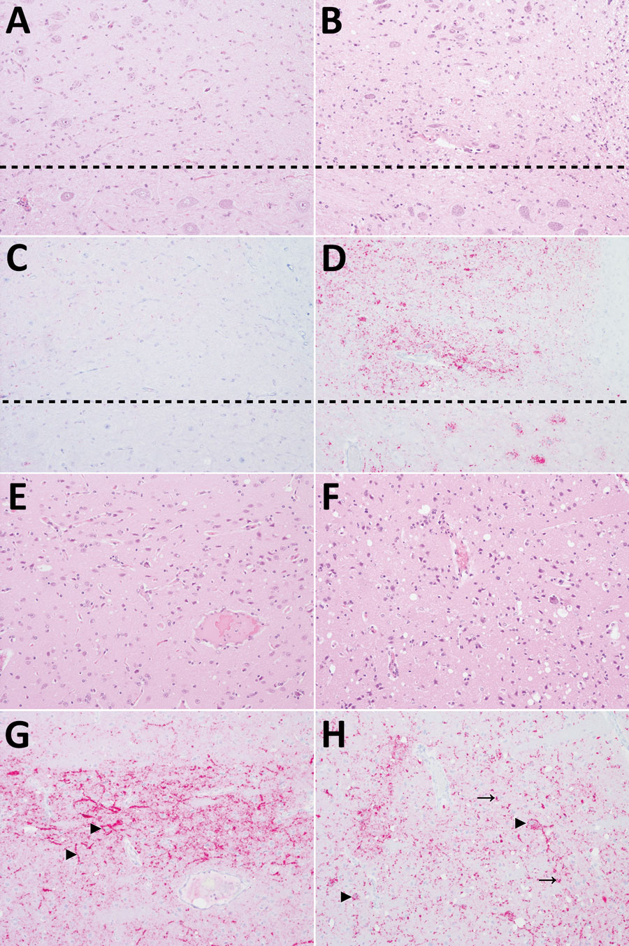
Figure 4. Patterns of histopathology and immunohistopathology in brains from 2 raccoons inoculated with the agent of chronic wasting disease (CWD). Panels A, C, E, and G show results for raccoon 2, inoculated with the agent of CWD from white-tailed deer; panels B, D, F, and H) show results for raccoon 9, inoculated with CWD from a vole that had been inoculated with the 4th vole-passage of the agent of CWD from white-tailed deer. All images original magnification ×20. A–D) Medulla at the level of the obex. A) Raccoon 2 shows no spongiform change in the dorsal motor nucleus of the vagus nerve (DMNV) (above dashed line) or hypoglossal nucleus (below dashed line). Hematoxylin and eosin (H&E) stain. B) Raccoon 9 shows mild to moderate spongiform change in the DMNV. H&E stain. C) Raccoon 2 shows very mild PrPSc immunoreactivity in the DMNV and no immunoreactivity in neurons of the hypoglossal nucleus. PrP antibodies F89/160.1.5 and F99/97.6.1. D) Raccoon 9 shows moderate PrPSc immunoreactivity in the neuropil of the DMNV and marked intraneuronal immunoreactivity in the hypoglossal nucleus. PrP antibodies F89/160.1.5 and F99/97.6.1. E–H) Caudate nucleus. E) Raccoon 2 shows moderate diffuse spongiform change. H&E stain. F) Raccoon 9 shows marked diffuse spongiform change. H&E stain. G) Raccoon 2 shows diffuse neuropil PrPSc immunoreactivity and prominent extracellular PrPSc accumulation on neurons (arrowheads). PrP antibodies F89/160.1.5 and F99/97.6.1. H) Raccoon 9 shows marked intracellular PrPSc immunoreactivity in neurons (arrowheads) and glial cells (arrows). PrP antibodies F89/160.1.5 and F99/97.6.1.