Volume 28, Number 4—April 2022
Research
Increased Attack Rates and Decreased Incubation Periods in Raccoons with Chronic Wasting Disease Passaged through Meadow Voles
Cite This Article
Citation for Media
Abstract
Chronic wasting disease (CWD) is a naturally-occurring neurodegenerative disease of cervids. Raccoons (Procyon lotor) and meadow voles (Microtus pennsylvanicus) have previously been shown to be susceptible to the CWD agent. To investigate the potential for transmission of the agent of CWD from white-tailed deer to voles and subsequently to raccoons, we intracranially inoculated raccoons with brain homogenate from a CWD-affected white-tailed deer (CWDWtd) or derivatives of this isolate after it had been passaged through voles 1 or 5 times. We found that passage of the CWDWtd isolate through voles led to a change in the biologic behavior of the CWD agent, including increased attack rates and decreased incubation periods in raccoons. A better understanding of the dynamics of cross-species transmission of CWD prions can provide insights into how these infectious proteins evolve in new hosts.
Transmissible spongiform encephalopathies, or prion diseases, are a group of fatal neurodegenerative diseases that include chronic wasting disease (CWD) in cervids, scrapie in sheep and goats, bovine spongiform encephalopathy (mad cow disease) in cattle, and Creutzfeldt-Jakob disease and Kuru in humans. As of January 2020, CWD has been reported in free-ranging and farmed cervids in 26 states in the United States and 3 provinces in Canada (1). CWD-affected cervids shed infectious prions into their environment during both the preclinical and clinical stages of disease (2–8), and infectivity persists in soil (9–13), on the surface of contaminated plant leaves and roots (14), and in association with mineral licks (15). Environmental contamination with CWD prions represents a source of infectious material to which noncervid wildlife species, including raccoons and other small mammals, can be exposed.
We previously reported the transmission of the agent of CWD from white-tailed deer (Odocoileus virginianus borealis) and elk to raccoons through experimental intracranial inoculation (16). Raccoons are able to propagate CWD prions from white-tailed deer and elk but with low attack rates (25%) and with disease-associated prion protein distribution restricted to the brain (16).
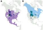
Successful transmission of the agent of CWD from white-tailed deer to 4 species of native North America rodents has been reported previously, and meadow voles (Microtus pennsylvanicus) were found to be the most susceptible species (17). Meadow voles are known to opportunistically scavenge carcasses and engage in cannibalistic behavior (18), providing a plausible route for exposure to CWD and the possibility of continued disease transmission. Small rodents are a food source for predators and scavengers, including raccoons, and meadow voles and raccoons inhabit overlapping geographic ranges that also overlap with locations undergoing cervid CWD epidemics (Figure 1). Therefore, the potential for direct exposure of meadow voles and raccoons to CWD infectivity in the environment exists. Indeed, studies in Wisconsin have shown that raccoons are present at deer carcasses and gut piles with a high frequency (19). In addition, because raccoons are mesopredators and scavengers, there is the potential for secondary exposure of raccoons through consumption of contaminated rodents.
To examine the potential for noncervid species to support CWD transmission, we intracranially inoculated raccoons with the agent of CWD from a white-tailed deer or with derivatives of the same inoculum after it had been passaged through meadow voles 1 or 5 times. In this study, we report the successful transmission of the agent of CWD from a white-tailed deer and vole-passaged CWD to raccoons through experimental intracranial inoculation. Our findings suggest passage of the CWD agent through voles results in a CWD agent with altered phenotypic properties.
We sourced 17 raccoon kits (8 weeks of age) that had no previous history of prion disease from a commercial breeder and challenged them by intracranial inoculation using 0.1 mL of a 10% brain homogenate (20). Brain material from 3 CWD-affected donor animals generated in a previous study (17) were used as inocula: 1 hunter-harvested (year of harvest 2001) CWD-positive white-tailed deer that was heterozygous for glycine and serine at codon 96 of the prion protein (GS96) (CWDWtd), 1 meadow vole that had been inoculated intracranially with the CWDWtd inoculum (first passage, CWDVole-P1), and 1 meadow vole that had been inoculated intracranially with brain material from a fourth passage vole (fifth passage, CWDVole-P5) (Table; Appendix). We inoculated raccoons in the negative control groups with brain material from a vole that had been intracranially inoculated with obex tissue from a CWD-negative deer (CWDNeg) (Table; Appendix). We prepared each inoculum from a single donor animal; no pooling was performed. We monitored raccoons daily and euthanized them when they showed unequivocal signs of prion disease (such as ataxia, inability to climb, or recumbency), when intercurrent illness or injury was present that could not be remedied by veterinary care, or at the end of the experiment at 35 months after inoculation. At raccoon death, we performed a full necropsy on all raccoons. We fixed 1 set of tissue samples in 10% buffered formalin, embedded in paraffin wax, and sectioned at 5 μm for microscopy examination after hematoxylin and eosin staining or immunohistochemical staining for detection of disease-associated prion protein (PrPSc) by using a cocktail containing 2 monoclonal antibodies, F89/160.1.5 and F99/97.6.1 (Appendix). We froze the second set of tissues, comprising subsamples of all tissues collected into formalin, and examined selected samples for the presence of disease-associated prion protein (PrPSc) by using a commercially available antigen-capture enzyme immunoassay or in-house Western blotting (Appendix).
Ethics Statement
This experiment was carried out in accordance with the Guide for the Care and Use of Laboratory Animals (Institute of Laboratory Animal Resources, National Academy of Sciences, Washington, DC, USA) and the Guide for the Care and Use of Agricultural Animals in Research and Teaching (Federation of Animal Science Societies, Champaign, IL, USA). The Institutional Animal Care and Use Committee at the National Animal Disease Center reviewed and approved the animal use protocols (approval no. ARS-2778).
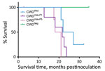
In the CWDWtd group, 3/4 raccoons demonstrated clinical signs consistent with prion disease (ataxia, inability to climb, recumbency); the average survival time was 23 months postinoculation (mpi) (Table). The remaining raccoon was euthanized at 32 mpi because of bilateral eye lesions; PrPSc was not detected in any tissues examined. We detected PrPSc in all raccoons in the CWDVole-P1 group. Two raccoons were euthanized or found dead because of urinary tract disease, and 1 was euthanized at 22 mpi because of lameness that was not responsive to treatment. The remaining 2 raccoons exhibited ataxia and inability to climb and were euthanized at 22 and 24 mpi (Table). In the CWDVole-P5 group, 2 raccoons were euthanized because of urinary tract disease at 3 mpi (PrPSc not detected) and 17 mpi (PrPSc-positive). During 18–21 mpi, the remaining 3 raccoons demonstrated ataxia and inability to climb; 2 of these animals also showed head tremors (Table). All 4 raccoons in the CWDNeg control group were clinically normal when they were euthanized at the end of the study at 35 mpi (Figure 2). By using antigen-capture enzyme immunoassay, we detected PrPSc in the brains of 3/4 raccoons in the CWDWtd group, 5/5 raccoons in the CWDVole-P1 group, 4/4 raccoons in the CWDVole-P5 group (not including the raccoon that was euthanized because of urinary tract disease at 3 mpi), and 0/4 raccoons in the CWDNeg group (Table).
When we analyzed brain samples by Western blot by using monoclonal PrP antibody P4, migration patterns for all animals within a treatment group were similar to each other and to the original inoculum (data not shown). When we compared samples across groups, migration patterns for vole-passaged groups were similar to each other with the unglycosylated band at ≈19 kDa. The unglycosylated band of the sample from the CWDWtd group migrated slightly higher, at 20 kDa, and that of the original donor white-tailed deer migrated slightly higher again, at ≈21 kDa (Figure 3).
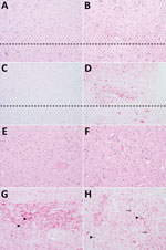
We examined hematoxylin and eosin–stained sections to assess pathologic changes in the brain (Figure 4). Immunohistochemical staining for PrPSc was applied to the brain and peripheral tissues to investigate the distribution of PrPSc throughout the body (Figure 4). In raccoons in the CWDWtd group, spongiform change of the neurophil was mild caudally (medulla at the level of the obex and midbrain) and moderate rostrally (thalamus and basal nuclei). Spongiform change was not observed in the dorsal motor nucleus of the vagus nerve (Figure 4, panel A) or cerebellum and was mild to moderate in the basal nuclei (Figure 4, panel E) and neocortex. In contrast, spongiform change in the vole-passaged CWD groups was moderate to marked throughout the brain, including in the dorsal motor nucleus of the vagus nerve (Figure 4, panel B), basal nuclei (Figure 4, panel F) and neocortex, and mild in the cerebellum. Intraneuronal vacuolation was only observed in 2 raccoons, both of which were from the vole-passaged CWD groups. A single intraneuronal vacuole was seen in the red nucleus of raccoon 6 (CWDVole-P1) and the dorsal motor nucleus of raccoon 14 (CWDVole-P5).
We detected immunoreactivity for PrPSc in the brain, spinal cord, retina, optic nerve, and/or pituitary in >2 raccoons per group (Table). We did not detect PrPSc in any lymphoid tissues sampled but was observed in the enteric nervous system of the stomach, jejunum, ileum and colon of raccoon 3 (CWDWtd).
In the brains of raccoons in the CWDWtd group, the overall amount of PrPSc immunoreactivity was less in the caudal parts of the brain (Figure 4, panel C) and greater in the rostral parts of the brain (thalamus and basal nuclei) (Figure 4, panel G). Extracellular PrPSc accumulation in the neuropil and on neurons was more prominent than intraneuronal accumulation (Figure 4, panels C, G). In contrast, the pattern of PrPSc immunoreactivity was similar in raccoons in the vole-passaged CWD groups and characterized by PrPSc immunoreactivity throughout the brain with intracellular PrPSc accumulation in microglia, astrocytes, and neurons (Figure 4, panels D, H).
To enable objective comparisons of the distribution and severity of spongiform change between inoculation groups, we scored the severity of vacuolation on a scale of 0–4 for 17 neuroanatomical areas and used the score to generate vacuolation lesion profiles as described previously (21). We made modifications to include the red nucleus and dorsal motor nucleus of the vagus nerve, which resulted in a total of 19 neuroanatomical areas examined. The distribution of vacuolation in raccoons was similar in the vole-passaged CWD groups, although the overall severity of vacuolation was greater in the CWDVole-P5 group than the CWDVole-P1 group (Figure 5). The pattern of vacuolation observed in raccoons in the CWDWtd group was different from raccoons in the vole-passaged groups. A trend for less severe vacuolation overall was particularly noticeable in the medulla (Figure 5, neuroanatomical areas 1–4), midbrain (Figure 5, areas 8–9), frontal cortex (Figure 5, area 13), and claustrum (Figure 5, area 17) (Appendix).
We demonstrated that clinical disease developed in raccoons inoculated intracranially with the agent of CWD from white-tailed deer (CWDWtd); the average incubation period was ≈23 mpi. Passage of the CWDWtd isolate through meadow voles before inoculation of raccoons with this vole-passaged CWD resulted in slightly shorter incubation periods (≈20 mpi) and different neuropathology and western blot migration pattern as compared with the original CWDWtd isolate.
We previously reported that experimental intracerebral inoculation of raccoons with an inoculum prepared from pooled brainstems from 11 CWD-affected white-tailed deer (CWDWtd-pool) resulted in disease in 1/4 raccoons with an incubation period of 73 mpi and restricted distribution of PrPSc accumulation in the brain (16). The low attack rate and prolonged incubation period produced by the CWDWtd-pool inoculum compared with the CWDWtd inoculum reported in this study could be due to differences in the titer of PrPSc in the donor inocula. However, we consider this scenario unlikely, because all donor deer used to prepare the CWDWtd-pool inoculum and the single donor deer used to prepare the CWDWtd inoculum were positive by immunohistochemistry for PrPSc. Another point of difference in disease expression produced by the CWDWtd-pool compared with the CWDWtd inoculum is the pattern of neuropathology observed in the brain: vacuolation of neuronal perikarya was widespread in the brain of the raccoon inoculated with the CWDWtd-pool (16) but was not observed in raccoons inoculated with CWDWtd. The differences in biologic behavior of these 2 CWD isolates are most likely associated with differences in the prion protein (PRNP) genotype of the donor deer. Four PRNP polymorphisms exist in white-tailed deer: Q95H, G96S, A116G, and Q226K (reviewed in S.J. Robinson et al. [22]). At codon 96, the S96 allele is associated with reduced CWD prevalence (23–26) and prolonged incubation periods (27). Donor deer for the CWDWtd-pool inoculum were all GG96 PRNP genotype (16), whereas the donor deer for the CWDWtd inoculum was GS96 PRNP genotype. We did not expect that inoculum containing the S96 allele would produce disease in raccoons more efficiently than inoculum exclusively containing the G96 allele. In addition, sequencing of PRNP from raccoons in a previous study showed that raccoons are homozygous for glycine at codon 96 (GG96) (S.J. Moore, unpub. data). Therefore, our results suggest that patterns of disease susceptibility associated with PRNP polymorphisms at codon 96 in CWD-affected white-tailed deer might not be a useful predictor of disease outcomes in intracranially inoculated raccoons. Further studies are under way to investigate the biologic behavior in raccoons of CWD from a single-source GG96 white-tailed deer.
Cross-species transmission of CWD isolates might result in a change in the biochemical properties of the disease-associated prion protein or the biologic behavior of the prion strain, such as adaptation to its host, or both (16,28–33). The pattern of PrPSc accumulation in the brain of the raccoon inoculated with CWDWtd-pool in our previous study (16) was similar to raccoons inoculated with the CWDWtd inoculum in this study and was characterized by prominent linear and perineuronal PrPSc accumulation, although this comparison is limited by the small number of animals available for examination.
Both vole-passaged CWD isolates produced similar disease phenotypes in raccoons with regards to incubation periods, western blot migration patterns, and neuropathology. The patterns of spongiform change and PrPSc accumulation in the brains of raccoons inoculated with vole-passaged CWD isolates were similar to each other and different from those observed in the brains of raccoons inoculated with the CWDWtd isolate. Inoculum-associated differences in western blot migration patterns were observed (i.e., similar patterns for vole-passaged CWD isolates and a different pattern for the CWDWtd isolate). In addition, the migration pattern of the original CWD-affected white-tailed deer donor (Figure 3, deer CWD, unglycosylated band at ≈21 kDa) was different from both the raccoon-passaged CWDWtd isolate (Figure 3, white-tailed deer, 20 kDa) and the vole-passaged CWD isolates (Figure 3, vole-P1 and vole-P5, 19 kDa) after passage through raccoons. Therefore, passage of CWDWtd through voles appears to result in a change in the biologic behavior of this prion isolate when inoculated intracranially into raccoons. This finding raises the possibility for emergence of novel CWD strains after passage in off-target species through host-driven selection of a strain present in the donor inoculum (29,34). The original inoculum was derived from a white-tailed deer with the GS96 PRNP genotype, and propogation of CWD prions on S96 PrPC results in the formation of alternative PrPSc conformers (34). We are unsure what role the genotype of the deer in the inoculum might have played in the change in biologic behavior noted after passage through voles. Because intracranial inoculation is not a natural route for exposure of raccoons to CWD infection, oral transmission studies are underway to characterize the biologic behavior of the CWDWtd and vole-passaged CWD isolates using a more natural route of exposure.
We observed a single intraneuronal vacuole in the red nucleus of raccoon 6 (CWDVole-P1 group) and the dorsal motor nucleus of raccoon 14 (CWDVole-P5 group). Intraneuronal vacuolation was previously reported as an incidental finding in the brainstem (including facial and pontine nuclei), cerebellar roof nuclei, and cerebrum of raccoons (35,36). In those raccoons, no evidence of concurrent neuropil vacuolation, neuronal degeneration, or astrocytosis was seen. In contrast, widespread neuropil vacuolation throughout the brains of raccoons 6 and 14, and strong PrPSc immunoreactivity in vacuolated neurons was evident; therefore, the intraneuronal vacuoles observed in these raccoons are likely associated with prion infection.
Although PrPSc immunoreactivity was widely distributed throughout the brain and spinal cord, we did not generally observe involvement of the peripheral nervous system, with the exception of 1 raccoon (3) inoculated with CWDWtd, in which PrPSc immunoreactivity was present in the enteric plexi of the stomach, jejunum, ileum, and colon. The general lack of peripheral nervous system involvement is likely because raccoons were inoculated through the intracranial route that bypasses centripetal spread of PrPSc from the alimentary tract to the brain along parasympathetic nerves, as is observed in orally infected deer (37). Instead, PrPSc immunoreactivity in the enteric nervous system of raccoon 3 was likely the result of centrifugal spread from the central nervous system. Why PrPSc immunoreactivity was observed throughout the spinal cord in all raccoons is unclear, but enteric nervous system involvement was only seen in raccoon 3. Raccoon 3 was the longest surviving raccoon (27 mpi), so the possibility exists that, had other raccoons not succumbed to clinical disease, there might have been time for transport of PrPSc to the enteric nervous system. Inoculation of raccoons by the oral route is needed to improve our understanding of the pathogenesis of CWD in raccoons when exposure occurs by a more natural route.
The longest surviving CWD-inoculated raccoon (4) was euthanized at 32 mpi because of bilateral eye lesions. Histopathologic examination resulted in a diagnosis of multicentric lymphoma, and PrPSc was not detected in any tissues. Clinical disease and widespread PrPSc accumulation at 21–27 mpi developed in all other raccoons in the CWDWtd group (n = 3). The reason for the unexpected negative result for raccoon 4 is unclear but could include experimental error or host factors. With regard to experimental inoculation, all inocula were prepared and all raccoons were inoculated on the same day, so the likelihood is very low that this raccoon did not receive the correct inoculum. The strongest determinant of susceptibility to prion diseases is the host PRNP sequence. No unique single nucleotide polymorphisms were detected in the PRNP open reading frame of raccoon 4 (S.J. Moore, unpub. data). It is tempting to speculate that host, genetic, or immunological factors outside of the PRNP open reading frame that contributed to the development of neoplasia might have had a suppressive effect on PrPSc accumulation.
Prion diseases of free-ranging animals do not exist in isolation. Meadow voles and raccoons are widespread in North America, and their habitat ranges overlap with those of CWD-affected white-tailed deer and other cervids. Therefore, a substantial potential for exposure of these or other off-target species to CWD infectivity in the environment exists. We have demonstrated that CWDWtd from a GS96 white-tailed deer transmitted readily to raccoons. Passage of this isolate through voles followed by intracranial inoculation of raccoons with vole-derived inoculum resulted in disease with different biologic characteristics and neuropathology than the original CWDWtd isolate. These results provide strong evidence for the emergence of a novel strain of CWD after passage in meadow voles and raccoons. Therefore, interspecies transmission of CWD prions between cervids and noncervid species that share the same habitat might represent a confounding factor in CWD-management programs. In addition, passage of CWD prions through off-target species might represent a source of novel CWD strains with unknown biologic characteristics, including zoonotic potential. Characterization of the biologic behavior of CWD isolates after cross-species transmission will help us develop more effective management strategies for CWD-affected populations.
Dr. Moore performed this work as a postdoctoral research associate at the National Animal Disease Center, US Department of Agriculture, Ames, Iowa. Her research interests include pathogenesis and pathology of animal diseases with a special interest in neuropathology and interspecies transmission of prion diseases.
Acknowledgments
We would like to thank Martha Church, Joe Lesan, Leisa Mandell, and Kevin Hassall for excellent technical support. Raccoon and vole range data were provided by NatureServe in collaboration with Bryan Richards, USGS National Wildlife Health Center, and range figures were generated by Andrew Fox (Centers for Epidemiology and Animal Health, Animal and Plant Health Inspection Service, US Department of Agriculture).
This research was supported in part by an appointment (S.J.M.) to the Agricultural Research Service Research Participation Program administered by the Oak Ridge Institute for Science and Education (ORISE) through an interagency agreement between the US Department of Energy (DOE) and the US Department of Agriculture. ORISE is managed by Oak Ridge Associated Universities (ORAU) under DOE contract no. DE-SC0014664.
References
- US Geological Survey National Wildlife Health Center. 2020 [cited 2020 Jan 18]. https://www.nwhc.usgs.gov/disease_information/chronic_wasting_disease/index.jsp
- Mathiason CK, Powers JG, Dahmes SJ, Osborn DA, Miller KV, Warren RJ, et al. Infectious prions in the saliva and blood of deer with chronic wasting disease. Science. 2006;314:133–6. DOIPubMedGoogle Scholar
- Mathiason CK, Hays SA, Powers J, Hayes-Klug J, Langenberg J, Dahmes SJ, et al. Infectious prions in pre-clinical deer and transmission of chronic wasting disease solely by environmental exposure. PLoS One. 2009;4:
e5916 . DOIPubMedGoogle Scholar - Haley NJ, Seelig DM, Zabel MD, Telling GC, Hoover EA. Detection of CWD prions in urine and saliva of deer by transgenic mouse bioassay. PLoS One. 2009;4:
e4848 . DOIPubMedGoogle Scholar - Henderson DM, Manca M, Haley NJ, Denkers ND, Nalls AV, Mathiason CK, et al. Rapid antemortem detection of CWD prions in deer saliva. PLoS One. 2013;8:
e74377 . DOIPubMedGoogle Scholar - Haley NJ, Mathiason CK, Zabel MD, Telling GC, Hoover EA. Detection of sub-clinical CWD infection in conventional test-negative deer long after oral exposure to urine and feces from CWD+ deer. PLoS One. 2009;4:
e7990 . DOIPubMedGoogle Scholar - Tamgüney G, Miller MW, Wolfe LL, Sirochman TM, Glidden DV, Palmer C, et al. Asymptomatic deer excrete infectious prions in faeces. [Erratum in: Nature. 2010;466:652]. Nature. 2009;461:529–32. DOIPubMedGoogle Scholar
- Pulford B, Spraker TR, Wyckoff AC, Meyerett C, Bender H, Ferguson A, et al. Detection of PrPCWD in feces from naturally exposed Rocky Mountain elk (Cervus elaphus nelsoni) using protein misfolding cyclic amplification. J Wildl Dis. 2012;48:425–34. DOIPubMedGoogle Scholar
- Johnson CJ, Phillips KE, Schramm PT, McKenzie D, Aiken JM, Pedersen JA. Prions adhere to soil minerals and remain infectious. PLoS Pathog. 2006;2:
e32 . DOIPubMedGoogle Scholar - Johnson CJ, Pedersen JA, Chappell RJ, McKenzie D, Aiken JM. Oral transmissibility of prion disease is enhanced by binding to soil particles. PLoS Pathog. 2007;3:
e93 . DOIPubMedGoogle Scholar - Seidel B, Thomzig A, Buschmann A, Groschup MH, Peters R, Beekes M, et al. Scrapie Agent (Strain 263K) can transmit disease via the oral route after persistence in soil over years. PLoS One. 2007;2:
e435 . DOIPubMedGoogle Scholar - Jacobson KH, Lee S, Somerville RA, McKenzie D, Benson CH, Pedersen JA. Transport of the pathogenic prion protein through soils. J Environ Qual. 2010;39:1145–52. DOIPubMedGoogle Scholar
- Miller MW, Williams ES, Hobbs NT, Wolfe LL. Environmental sources of prion transmission in mule deer. Emerg Infect Dis. 2004;10:1003–6. DOIPubMedGoogle Scholar
- Pritzkow S, Morales R, Moda F, Khan U, Telling GC, Hoover E, et al. Grass plants bind, retain, uptake, and transport infectious prions. Cell Rep. 2015;11:1168–75. DOIPubMedGoogle Scholar
- Plummer IH, Johnson CJ, Chesney AR, Pedersen JA, Samuel MD. Mineral licks as environmental reservoirs of chronic wasting disease prions. PLoS One. 2018;13:
e0196745 . DOIPubMedGoogle Scholar - Moore SJ, Smith JD, Richt JA, Greenlee JJ. Raccoons accumulate PrPSc after intracranial inoculation of the agents of chronic wasting disease or transmissible mink encephalopathy but not atypical scrapie. J Vet Diagn Invest. 2019;31:200–9. DOIPubMedGoogle Scholar
- Heisey DM, Mickelsen NA, Schneider JR, Johnson CJ, Johnson CJ, Langenberg JA, et al. Chronic wasting disease (CWD) susceptibility of several North American rodents that are sympatric with cervid CWD epidemics. J Virol. 2010;84:210–5. DOIPubMedGoogle Scholar
- Zajac AM, Williams JF. The pathology of infection with Schistosomatium douthitti in the laboratory mouse and the meadow vole, Microtus pennsylvanicus. J Comp Pathol. 1981;91:1–10. DOIPubMedGoogle Scholar
- Jennelle CS, Samuel MD, Nolden CA, Berkley EA. Deer carcass decomposition and potential scavenger exposure to chronic wasting disease. J Wildl Manage. 2009;73:655–62. DOIGoogle Scholar
- Hamir AN, Miller JM, Cutlip RC, Stack MJ, Chaplin MJ, Jenny AL, et al. Experimental inoculation of scrapie and chronic wasting disease agents in raccoons (Procyon lotor). Vet Rec. 2003;153:121–3. DOIPubMedGoogle Scholar
- Simmons MM, Harris P, Jeffrey M, Meek SC, Blamire IW, Wells GA. BSE in Great Britain: consistency of the neurohistopathological findings in two random annual samples of clinically suspect cases. Vet Rec. 1996;138:175–7. DOIPubMedGoogle Scholar
- Robinson SJ, Samuel MD, O’Rourke KI, Johnson CJ. The role of genetics in chronic wasting disease of North American cervids. Prion. 2012;6:153–62. DOIPubMedGoogle Scholar
- Robinson SJ, Samuel MD, Johnson CJ, Adams M, McKenzie DI. Emerging prion disease drives host selection in a wildlife population. Ecol Appl. 2012;22:1050–9. DOIPubMedGoogle Scholar
- Johnson C, Johnson J, Vanderloo JP, Keane D, Aiken JM, McKenzie D. Prion protein polymorphisms in white-tailed deer influence susceptibility to chronic wasting disease. J Gen Virol. 2006;87:2109–14. DOIPubMedGoogle Scholar
- O’Rourke KI, Spraker TR, Hamburg LK, Besser TE, Brayton KA, Knowles DP. Polymorphisms in the prion precursor functional gene but not the pseudogene are associated with susceptibility to chronic wasting disease in white-tailed deer. J Gen Virol. 2004;85:1339–46. DOIPubMedGoogle Scholar
- Johnson C, Johnson J, Clayton M, McKenzie D, Aiken J. Prion protein gene heterogeneity in free-ranging white-tailed deer within the chronic wasting disease affected region of Wisconsin. J Wildl Dis. 2003;39:576–81. DOIPubMedGoogle Scholar
- Johnson CJ, Herbst A, Duque-Velasquez C, Vanderloo JP, Bochsler P, Chappell R, et al. Prion protein polymorphisms affect chronic wasting disease progression. PLoS One. 2011;6:
e17450 . DOIPubMedGoogle Scholar - Angers RC, Kang HE, Napier D, Browning S, Seward T, Mathiason C, et al. Prion strain mutation determined by prion protein conformational compatibility and primary structure. Science. 2010;328:1154–8. DOIPubMedGoogle Scholar
- Duque Velásquez C, Kim C, Herbst A, Daude N, Garza MC, Wille H, et al. Deer prion proteins modulate the emergence and adaptation of chronic wasting disease strains. J Virol. 2015;89:12362–73. DOIPubMedGoogle Scholar
- Perrott MR, Sigurdson CJ, Mason GL, Hoover EA. Evidence for distinct chronic wasting disease (CWD) strains in experimental CWD in ferrets. J Gen Virol. 2012;93:212–21. DOIPubMedGoogle Scholar
- Raymond GJ, Raymond LD, Meade-White KD, Hughson AG, Favara C, Gardner D, et al. Transmission and adaptation of chronic wasting disease to hamsters and transgenic mice: evidence for strains. J Virol. 2007;81:4305–14. DOIPubMedGoogle Scholar
- Aguilar-Calvo P, Bett C, Sevillano AM, Kurt TD, Lawrence J, Soldau K, et al. Generation of novel neuroinvasive prions following intravenous challenge. Brain Pathol. 2018;28:999–1011. DOIPubMedGoogle Scholar
- Herbst A, Velásquez CD, Triscott E, Aiken JM, McKenzie D. Chronic wasting disease prion strain emergence and host range expansion. Emerg Infect Dis. 2017;23:1598–600. DOIPubMedGoogle Scholar
- Duque Velásquez C, Kim C, Haldiman T, Kim C, Herbst A, Aiken J, et al. Chronic wasting disease (CWD) prion strains evolve via adaptive diversification of conformers in hosts expressing prion protein polymorphisms. J Biol Chem. 2020;295:4985–5001. DOIPubMedGoogle Scholar
- Hamir AN, Fischer KA. Neuronal vacuolation in raccoons from Oregon. J Vet Diagn Invest. 1999;11:303–7. DOIPubMedGoogle Scholar
- Hamir AN, Heidel JR, Picton R, Rupprecht CE. Neuronal vacuolation in raccoons (Procyon lotor). Vet Pathol. 1997;34:250–2. DOIPubMedGoogle Scholar
- Fox KA, Jewell JE, Williams ES, Miller MW. Patterns of PrPCWD accumulation during the course of chronic wasting disease infection in orally inoculated mule deer (Odocoileus hemionus). J Gen Virol. 2006;87:3451–61. DOIPubMedGoogle Scholar
Figures
Cite This ArticleOriginal Publication Date: March 07, 2022
Table of Contents – Volume 28, Number 4—April 2022
| EID Search Options |
|---|
|
|
|
|
|
|


Please use the form below to submit correspondence to the authors or contact them at the following address:
Justin Greenlee, National Animal Disease Center, ARS, USDA, 1920 Dayton Ave, PO Box 70, Ames, IA 50010, USA
Top