Volume 28, Number 4—April 2022
Dispatch
Rigidoporus corticola Colonization and Invasive Fungal Disease in Immunocompromised Patients, United States
Figure 2

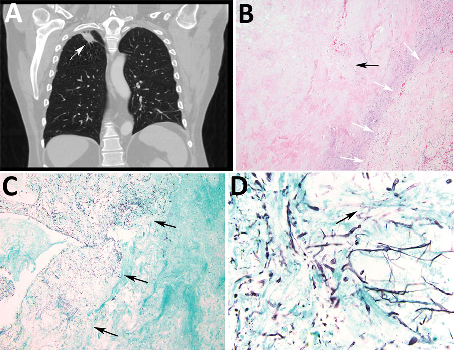
Figure 2. Radiologic and pathologic findings in a 61-year-old immunocompromised man with a history of lung adenocarcinoma and a new 2.4 cm right upper lobe mass determined to be a pulmonary fungus ball caused by Rigidoporus corticola (Oxyporus corticola) infection, United States. A) Chest computed tomography scan. Arrow indicates a 2.4 × 2.2 cm ovoid pulmonary mass in the right upper lobe. B) Hematoxylin and eosin–stained histologic sections of the resected mass. Black arrow indicates a cavitary lesion with a necrotic center. White arrows indicate peripheral fibrous capsule. Original magnification ×100. C, D) Gomori methenamine silver–stained histologic sections. C) Arrows indicate numerous hyphae within the cavity, but no evident invasion into blood vessels or surrounding tissue. Original magnification ×40. D) Arrow indicates septate thin hyphae evident in the center of the cavity. Original magnification ×600.