Volume 28, Number 4—April 2022
Research Letter
Autochthonous Leishmania infantum in Dogs, Zambia, 2021
David Squarre1, Herman M. Chambaro1 , Kyoko Hayashida1, Lavel C. Moonga, Yongjin Qiu, Yasuyuki Goto, Elizabeth Oparaocha, Chisoni Mumba, Walter Muleya, Patricia Bwalya, Joseph Chizimu, Mwelwa Chembensofu, Edgar Simulundu, Wizaso Mwasinga, Nelly Banda, Racheal Mwenda, Junya Yamagishi, King S. Nalubamba, Fredrick Banda, Musso Munyeme, Hirofumi Sawa, and Paul Fandamu
Figure
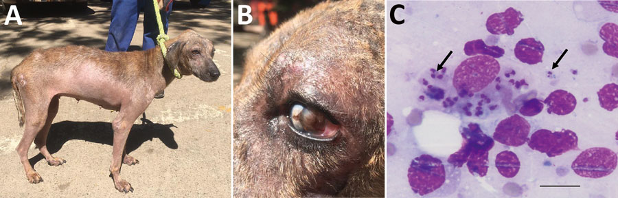
Figure. Clinical manifestations and microscopic imaging of Leishmania infantum‒infected dog, Zambia. A, B) Dog (case 1) showing dermatitis and onychogryphosis (excessive growth of nails) (A) and focal corneal opacity of the left eye (B). C) Intracellular Leishmania amastigotes (black arrows) in fine-needle lymph node aspirate from the same dog. Scale bar indicates 20 μm.
1These authors contributed equally to this article
Page created: December 22, 2021
Page updated: March 19, 2022
Page reviewed: March 19, 2022
The conclusions, findings, and opinions expressed by authors contributing to this journal do not necessarily reflect the official position of the U.S. Department of Health and Human Services, the Public Health Service, the Centers for Disease Control and Prevention, or the authors' affiliated institutions. Use of trade names is for identification only and does not imply endorsement by any of the groups named above.