Volume 28, Number 5—May 2022
Research Letter
atpE Mutation in Mycobacterium tuberculosis Not Always Predictive of Bedaquiline Treatment Failure
Cite This Article
Citation for Media
Abstract
We report the emergence of an atpE mutation in a clinical Mycobacterium tuberculosis strain. Genotypic and phenotypic bedaquiline susceptibility testing displayed variable results over time and ultimately were not predictive of treatment outcome. This observation highlights the limits of current genotypic and phenotypic methods for detection of bedaquiline resistance.
Bedaquiline is one of the core drugs used to treat multidrug-resistant (MDR) tuberculosis (TB) and extensively drug-resistant TB (XDR TB) (1). Bedaquiline resistance is now part of the revised definition of XDR TB, and its incidence is rising alarmingly (2,3). Resistance to bedaquiline is mainly caused by mutations in Rv0678 (mmpR), which encodes the repressor of the efflux pump MmpL5−MmpS5, usually leading to low-level resistance (4). Conversely, mutations in atpE, which encodes the target of bedaquiline, the c subunit of the ATP synthase, are rarely described in clinical strains (5) and are associated with high increase of MICs (4). Mutations in pepQ and Rv1979c are also reported, but their effect on bedaquiline susceptibility is unclear. We report a case of an atpE mutation in a bedaquiline-resistant clinical strain of Mybobacterium tuberculosis and discuss the performances of current methods for susceptibility testing (Appendix) and their clinical implications (6).
A 32-year-old man from Georgia received a diagnosis of bilateral cavitary lung MDR TB upon his arrival in France in January 2020. Three consecutive treatment regimens of bedaquiline and clofazimine had failed. A fourth regimen combining bedaquiline, linezolid, cycloserine, clofazimine, delamanid, and amoxicillin/clavulanate + meropenem was initiated on arrival.
Figure
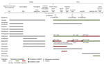
Figure. Timeline of treatment regimen and microbiologic data for patient with multidrug-resistant tuberculosis before and after his arrival in France from Georgia. Timeline for each antibiotic indicates treatment (striped), phenotypic resistance...
The first isolate from January 2020 (S1) was bedaquiline resistant with a MIC dilution above the breakpoint (MIC 2 mg/L) and clofazimine-susceptible with a MIC close to the breakpoint (MIC 1 mg/L). We detected 2 deletions (P129fs [15%] and G66fs [54%]) in Rv0678 (Figure).
At the end of March 2020, cycloserine was withdrawn from the patient’s treatment regimen because of phenotypic resistance, and bedaquiline, which had been stopped 1 month earlier, was resumed; the patient underwent a lobectomy. One month after the procedure, sputum microscopic examination and culture were still positive. The second isolate (S2) from April 2020 had an increased bedaquiline MIC (4 mg/L), but clofazimine MIC remained unchanged (1 mg/L). No mutation in Rv0678 was detected, but we observed an AtpE I66M (63%) substitution.
Two months later in June, the patient was sputum smear negative but remained culture positive. Isolate S3 was susceptible to bedaquiline (MIC 0.5 mg/L) and clofazimine resistant (MIC 2 mg/L). A deletion was found in Rv0678 different from those identified in S1: deletion at position 293 (N98fs) (97%), whereas no mutation was identified in atpE. Verapamil and ethionamide were added and amoxicillin/clavulanate + meropenem stopped. Finally, samples from September 2020 were culture negative, with regression of pulmonary lesions. The outcome was classified as treatment success in February 2021 after 13 months of treatment and was still favorable as of December 2021.
All 3 isolates shared the same spoligotype (SIT1) (Beijing lineage) and displayed only 3 single-nucleotide variants (SNVs) of difference by pairwise comparison. The SNVs were all nonsynonymous. Two SNVs were only recovered in strain S2, 1 corresponding to the AtpE: I66M substitution and 1 located in Rv0243 (L136P substitution) encoding the acetyl−CoA acyltransferase FadA2 and probably implicated in lipid degradation. One SNV was only found in S1 in Rv3909 (M683L substitution), encoding a protein of unknown function. No mutations were observed in pepQ, its promoter, or in Rv1979c (7).
As this case illustrates, identifying bedaquiline resistance in the laboratory and its effects on patient management appear complex. Over a 6-month period, we tested 3 M. tuberculosis isolates with different genotypic and phenotypic patterns regarding bedaquiline, exhibiting various MIC levels and mutations in genes involved in bedaquiline resistance. These isolates displayed only 3 SNVs by pairwise comparison of their genomes, excluding a reinfection by a new strain or a mixed infection.
Of note, mutations in atpE or Rv0678 were found only once and were not found at subsequent timepoints. Despite continuous bedaquiline treatment, resistant strain S2 with the atpE mutation was not selected, and the patient was cured. A previous in vitro study suggested that, whereas Rv0678 mutations were dynamic over time, atpE mutations were fixed once they appeared (8). This observation was not confirmed by our clinical case. One possible explanation for nonfixation of these mutations could be the associated fitness cost. However, an in vitro study did not show any fitness cost because of the I66M substitution (9). Because fitness also depends on the genetic background, the results of this in vitro study might not be transposable here. Regarding Rv0678, 2 mutations have been studied and did not have fitness impact (E138G and R94Q) (4). Additional in vivo and epidemiologic studies would help evaluate the fitness cost of such mutations. Another explanation for the variability of genotypic and phenotypic bedaquiline susceptibility over time could be a spatial heterogeneity in the lesions as already described (10).
This case raises concerns about the ability of current phenotypic and genotypic methods to detect bedaquiline resistance. Further studies are needed before relying on these methods for therapeutic decisions. In the meantime, these data can help improve the World Health Organization database of drug resistance–related mutations (11). Overall, this case underlines the complexity of bedaquiline-resistance mechanisms and of the dynamics of mutation emergence and selection.
Dr. Fournier Le Ray recently successfully defended a PhD thesis in microbiology. Her research was focused on the in vivo emergence of bedaquiline resistance.
Acknowledgments
We thank the technicians of NRC MyRMA for their technical assistance.
We wish to honor the memory of our colleague, Wladimir Sougakoff, who passed away in January 2022.
NRC MyRMA is supported by annual grant of Santé Publique France. L.F.L.R. was supported by a grant for Fondation du Souffle. The laboratory has received a research grant from Janssen for study on bedaquiline antimycobacterial activity.
References
- Van Deun A, Decroo T, Piubello A, de Jong BC, Lynen L, Rieder HL. Principles for constructing a tuberculosis treatment regimen: the role and definition of core and companion drugs. Int J Tuberc Lung Dis. 2018;22:239–45. DOIPubMedGoogle Scholar
- World Health Organization. Meeting report of the WHO expert consultation on the definition of extensively drug-resistant tuberculosis, 27–29 October 2020 [cited 2021 Feb 5]. https://www.who.int/publications/i/item/meeting-report-of-the-who-expert-consultation-on-the-definition-of-extensively-drug-resistant-tuberculosis
- Andres S, Merker M, Heyckendorf J, Kalsdorf B, Rumetshofer R, Indra A, et al. Bedaquiline-resistant tuberculosis: dark clouds on the horizon. Am J Respir Crit Care Med. 2020;201:1564–8. DOIPubMedGoogle Scholar
- Andries K, Villellas C, Coeck N, Thys K, Gevers T, Vranckx L, et al. Acquired resistance of Mycobacterium tuberculosis to bedaquiline. PLoS One. 2014;9:
e102135 . DOIPubMedGoogle Scholar - Peretokina IV, Krylova LY, Antonova OV, Kholina MS, Kulagina EV, Nosova EY, et al. Reduced susceptibility and resistance to bedaquiline in clinical M. tuberculosis isolates. J Infect. 2020;80:527–35. DOIPubMedGoogle Scholar
- World Health Organization. Technical manual for drug susceptibility testing of medicines used in the treatment of tuberculosis. 2018 [cited 2018 Oct 18]. https://apps.who.int/iris/handle/10665/275469
- Battaglia S, Spitaleri A, Cabibbe AM, Meehan CJ, Utpatel C, Ismail N, et al. Characterization of genomic variants associated with resistance to bedaquiline and delamanid in naive mycobacterium tuberculosis clinical strains. J Clin Microbiol. 2020;58:1304–24. DOIPubMedGoogle Scholar
- Ismail N, Ismail NA, Omar SV, Peters RPH. In vitro study of stepwise acquisition of rv0678 and atpE mutations conferring bedaquiline resistance. Antimicrob Agents Chemother. 2019;63:e00292–19. DOIPubMedGoogle Scholar
- Huitric E, Verhasselt P, Koul A, Andries K, Hoffner S, Andersson DI. Rates and mechanisms of resistance development in Mycobacterium tuberculosis to a novel diarylquinoline ATP synthase inhibitor. Antimicrob Agents Chemother. 2010;54:1022–8. DOIPubMedGoogle Scholar
- Kaplan G, Post FA, Moreira AL, Wainwright H, Kreiswirth BN, Tanverdi M, et al. Mycobacterium tuberculosis growth at the cavity surface: a microenvironment with failed immunity. Infect Immun. 2003;71:7099–108. DOIPubMedGoogle Scholar
- World Health Organization. Catalogue of mutations in Mycobacterium tuberculosis complex and their association with drug resistance [cited 2022 Jan 18]. https://www.who.int/publications-detail-redirect/9789240028173
Figure
Cite This ArticleOriginal Publication Date: April 11, 2022
Table of Contents – Volume 28, Number 5—May 2022
| EID Search Options |
|---|
|
|
|
|
|
|
Please use the form below to submit correspondence to the authors or contact them at the following address:
Nicolas Veziris, Faculté de Médecine Sorbonne Université, 91, boulevard de l’hôpital, 75013 Paris, France
Top