Volume 28, Number 7—July 2022
Research Letter
Hodgkin Lymphoma after Disseminated Mycobacterium genavense Infection, Germany
Figure
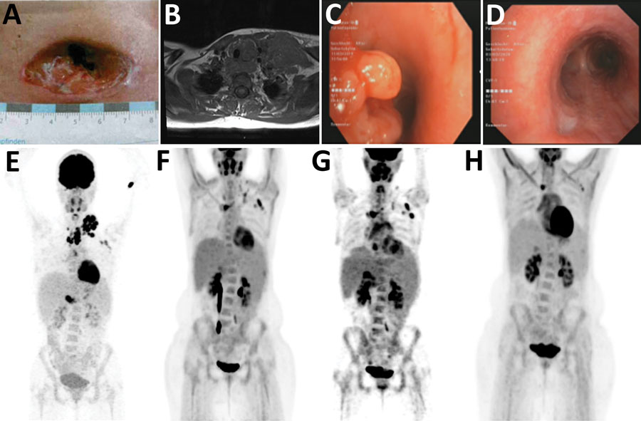
Figure. Clinical manifestations and radiologic findings in the course of disease in a 23-year-old woman with disseminated M. genavense infection preceding Hodgkin lymphoma, Germany. A) Cervical wound after initial lymph node extirpation. B) Magnetic resonance imaging at the time of initial evaluation. C) Endobronchial view of tracheo-esophageal fistula before positioning of a stent. D) Endobronchial view of the prior tracheo-esophageal fistula after treatment. Whitish scar tissue is seen at the bottom left. E) 18F-FDG-PET scan at initial evaluation (maximum intensity projection). Cervical lymph node mass is seen, with no pathologic uptake in the abdomen. F) 18F-FDG-PET scan after 6 months of antibiotic treatment showing reduced uptake. G) 18F-FDG-PET scan shortly before Hodgkin lymphoma was diagnosed showing new hepatosplenomegaly and lymphadenopathy. H) 18F-FDG-PET scan after antibiotic and chemotherapy without pathologic enhancement.