Volume 28, Number 8—August 2022
Dispatch
Novel Chronic Anaplasmosis in Splenectomized Patient, Amazon Rainforest
Olivier Duron , Rachid Koual, Lise Musset, Marie Buysse, Yann Lambert, Benoît Jaulhac, Denis Blanchet, Kinan Drak Alsibai, Yassamine Lazrek, Loïc Epelboin, Pierre Deshuillers, Céline Michaud, and Maylis Douine
Figure 2
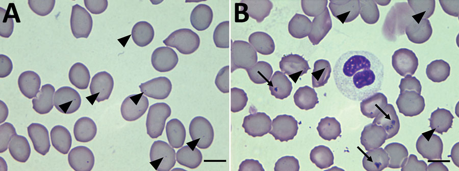
Figure 2. Thin films of a blood sample collected in October 2019 from a patient in French Guiana. Inclusions of Candidatus Anaplasma sparouinense are located at the periphery of the red blood cells as small round dots of 0.3–0.4 µm (arrowheads). Other red blood cells contain Howell-Jolly bodies of various shapes and sizes >1 µm (arrows). Some Howell-Jolly bodies are found in the background of the smears. Wright-Giemsa stain; original magnification ×100.
Page created: June 09, 2022
Page updated: July 20, 2022
Page reviewed: July 20, 2022
The conclusions, findings, and opinions expressed by authors contributing to this journal do not necessarily reflect the official position of the U.S. Department of Health and Human Services, the Public Health Service, the Centers for Disease Control and Prevention, or the authors' affiliated institutions. Use of trade names is for identification only and does not imply endorsement by any of the groups named above.