Volume 29, Number 1—January 2023
Dispatch
Photobacterium damselae subspecies damselae Pneumonia in Dead, Stranded Bottlenose Dolphin, Eastern Mediterranean Sea
Figure 2
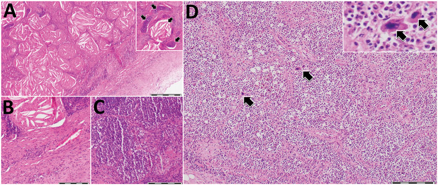
Figure 2. Histologic analysis of lungs and spleen of a bottlenose dolphin (Tursiops truncatus) with Photobacterium damselae subspecies damselae pneumonia, eastern Mediterranean Sea. A) Lung tissue showed a nodular structure covered by fibrous capsule (right bottom of figure panel) composed of numerous cholesterol clefts and areas of reactive fibrosis. Hyaline cartilage was observed, interpreted as bronchi and bronchioles. Inset, higher magnification showing an aggregate of cholesterol clefts and hyaline cartilage (arrow). B) Abundant fibrous lung tissue (lower right half) and cellular infiltrates were also observed. C) Different area of the lung parenchyma characterized by increased cellular infiltration. D) Spleen expressed an apparent contraction of the parenchyma showing diffuse cellularity, with only a few defined lymphoid follicles, as well as megakaryocytes (arrows) indicative of extramedullary hematopoiesis. Inset: higher magnification showing 2 adjacent megakaryocytes (arrows). Hematoxylin and eosin stained. Scale bars indicate 500 μm in panel A and 200 μm in panels B–D.