Skip directly to site content Skip directly to page options Skip directly to A-Z link Skip directly to A-Z link Skip directly to A-Z link
Volume 29, Number 10—October 2023
Dispatch

Influenza A(H5N1) Virus Infections in 2 Free-Ranging Black Bears (Ursus americanus), Quebec, Canada

Benjamin T. Jakobek, Yohannes Berhane, Marie-Soleil Nadeau, Carissa Embury-Hyatt, Oliver Lung, Wanhong Xu, and Stéphane LairComments to Author 
Author affiliations: Centre Québécois sur la Santé des Animaux Sauvages/Canadian Wildlife Health Cooperative, St. Hyacinthe, Quebec, Canada (B.T. Jakobek, S. Lair); Université de Montréal, St. Hyacinthe (B.T. Jakobek, S. Lair); University of Saskatchewan Western College of Veterinary Medicine, Saskatoon, Saskatchewan, Canada (Y. Berhane); Canadian Food Inspection Agency National Centre for Foreign Animal Disease, Winnipeg, Manitoba, Canada (Y. Berhane, C. Embury-Hyatt, O. Lung, W. Xu); University of Manitoba, Winnipeg (Y. Berhane, O. Lung); Ministère de l’Agriculture, des Pêcheries et de l’Alimentation Laboratoire de Santé Animale, St. Hyacinthe (M.-S. Nadeau)

Main Article

Figure 1

Detection of influenza A virus antigen in black bears by immunohistochemical analysis, Quebec, Canada. A) Brain tissue, showing abundant viral antigen detected multifocally throughout the section and observed primarily in gray matter areas. Scale bar indicates 5 mm. B) Brain immunostaining within neurons and surrounding neuropil. Scale bar indicates 100 μm. C) Liver tissue, showing viral antigen within individual cells. Scale bar indicates 200 μm. D) Liver tissue, showing that cells have the morphologic appearance of Kuppfer cells. Scale bar indicates 50 μm. Monoclonal antibody and diaminobenzidine stained, Gill’s hematoxylin counterstained.

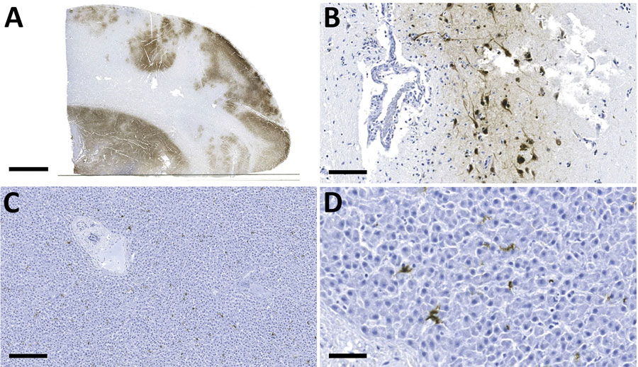
Figure 1. Detection of influenza A virus antigen in black bears by immunohistochemical analysis, Quebec, Canada. A) Brain tissue, showing abundant viral antigen detected multifocally throughout the section and observed primarily in gray matter areas. Scale bar indicates 5 mm. B) Brain immunostaining within neurons and surrounding neuropil. Scale bar indicates 100 μm. C) Liver tissue, showing viral antigen within individual cells. Scale bar indicates 200 μm. D) Liver tissue, showing that cells have the morphologic appearance of Kuppfer cells. Scale bar indicates 50 μm. Monoclonal antibody and diaminobenzidine stained, Gill’s hematoxylin counterstained.

Main Article

Page created: August 15, 2023
Page updated: September 20, 2023
Page reviewed: September 20, 2023
The conclusions, findings, and opinions expressed by authors contributing to this journal do not necessarily reflect the official position of the U.S. Department of Health and Human Services, the Public Health Service, the Centers for Disease Control and Prevention, or the authors' affiliated institutions. Use of trade names is for identification only and does not imply endorsement by any of the groups named above.
file_external